Duodenal-Type Follicular Lymphoma Mimic Follicular Hyperplasia
Keywords
Primary follicular lymphoma of duodenum, Spontaneous regression
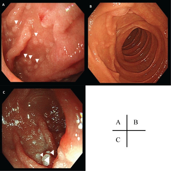
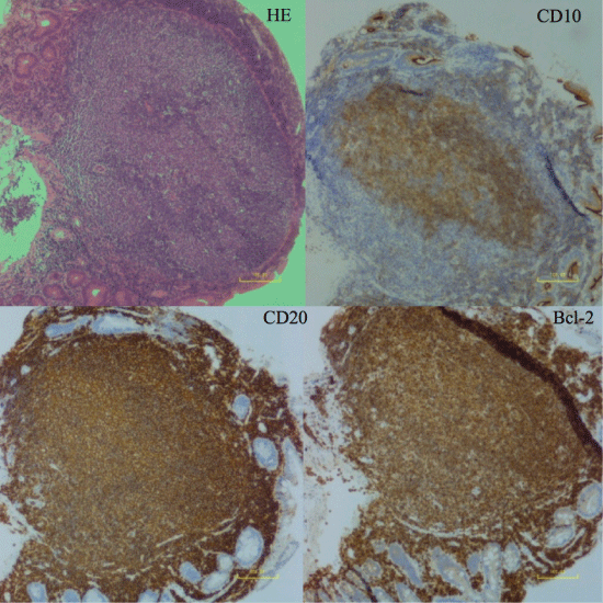
A 56-year-old Japanese woman with no notable medical history was admitted to our clinic because of abnormal changes in upper gastrointestinal series for regular checkup. An upper gastrointestinal endoscopy was performed. In the duodenum, white plaques resembling lymphoid hyperplasia were detected (Figure 1A, arrowheads). A biopsy was followed by the diagnosis of duodenal-type follicular lymphoma (FL) (Grade 1) (Figure 2). No other lesions were detected by further examinations (Stage I). Four courses of rituximab monotherapy (375 mg/m2 every 4 weeks) were administered. No antibiotics were used. At 1 year after the diagnosis, an upper endoscopy revealed the same lesion with slight regression. Two years after the diagnosis, the lesion had diminished (Figure 1B). Four years post-diagnosis, the lesion had reappeared but a biopsy revealed only lymphoid hyperplasia with no malignancy (Figure 1C, arrowheads). The patient is being followed up without any additional treatment.
Duodenal-type FL is a rare distinct variant of follicular disease characterized by lymphomatous disease limited to the duodenum, defined in the 2016 WHO classification [1,2]. Usually, multiple 1-5 mm polypoid lesions in the descending part of the duodenum are observed [1,3,4]. It is often difficult to distinguish duodenal polypoid lesions from lymphoid hyperplasia by endoscopic appearance, particularly when they are smaller in size or fewer in number. The most important thing is whether the gastroenterologist considers the possibility of FL and performs a biopsy or not. A pathological diagnosis is necessary to confirm the diagnosis.
Duodenal-type FL is distinct from other gastrointestinal-tract FLs. Duodenal-type FL patients appear to have an excellent prognosis, and watchful waiting and single-agent rituximab treatment may be sufficient [1,3,4].
Conflicts of Interest
The authors state that they have no conflicts of interest to declare.
References
- Schmatz A-I, Streubel B, Kretschmer-Chott E, et al. (2011) Primary follicular lymphoma of the duodenum is a distinct mucosal/submucosal variant of follicular lymphoma: a retrospective study of 63 cases. J Clin Oncol 29: 1445-1451.
- Swerdlow SH, Campo E, Pileri SA, et al. (2016) The 2016 revision of the World Health Organization classification of lymphoid neoplasms. Blood 127: 2375-2390.
- Shia J, Teruya-Feldstein J, Pan D, et al. (2002) Primary follicular lymphoma of the gastrointestinal tract: a clinical and pathologic study of 26 cases. Am J Surg Pathol 26: 216-224.
- Takata K, Okada H, Ohmiya N, et al. (2011) Primary gastrointestinal follicular lymphoma involving the duodenal second portion is a distinct entity: a multicenter, retrospective analysis in Japan. Cancer Sci 102: 1532-1536.
Corresponding Author
Dr. Yutaka Shimazu, Department of Hematology and Oncology, Graduate School of Medicine, Kyoto University, 54 Shogoin Kawahara-cho, Sakyo-ku, Kyoto 606-8507, Japan; Department of Hematology, Yamato Koriyama Hospital, Nara 639-1013, Japan, Tel: +81-75-751-4964, Fax: +81-75-751-4963.
Copyright
© 2017 Shimazu Y, et al. This is an open-access article distributed under the terms of the Creative Commons Attribution License, which permits unrestricted use, distribution, and reproduction in any medium, provided the original author and source are credited.


