Peripherally Inserted Central Catheter Malpositioned in the Ipsilateral Thoracodorsal Vein: Clinical Case
Clinical Case
A peripherally inserted central catheter (PICC) is a catheter that is inserted into a peripheral vein of the elbow or upper arm and which access the superior vena cava or inferior vena cava. These catheters present a low risk of serious complications but malpositioning of the distal tip is very frequent, with an incidence ranging from 5 to 31%. The most frequent tip locations of malpositioned PICC lines are the internal jugular vein, subclavian vein, axillary vein, brachial vein, contralateral veins, right atrium and right ventricle. Less frequently, the tip of the catheter may be placed in different arm veins and other peripheral veins [1-3].
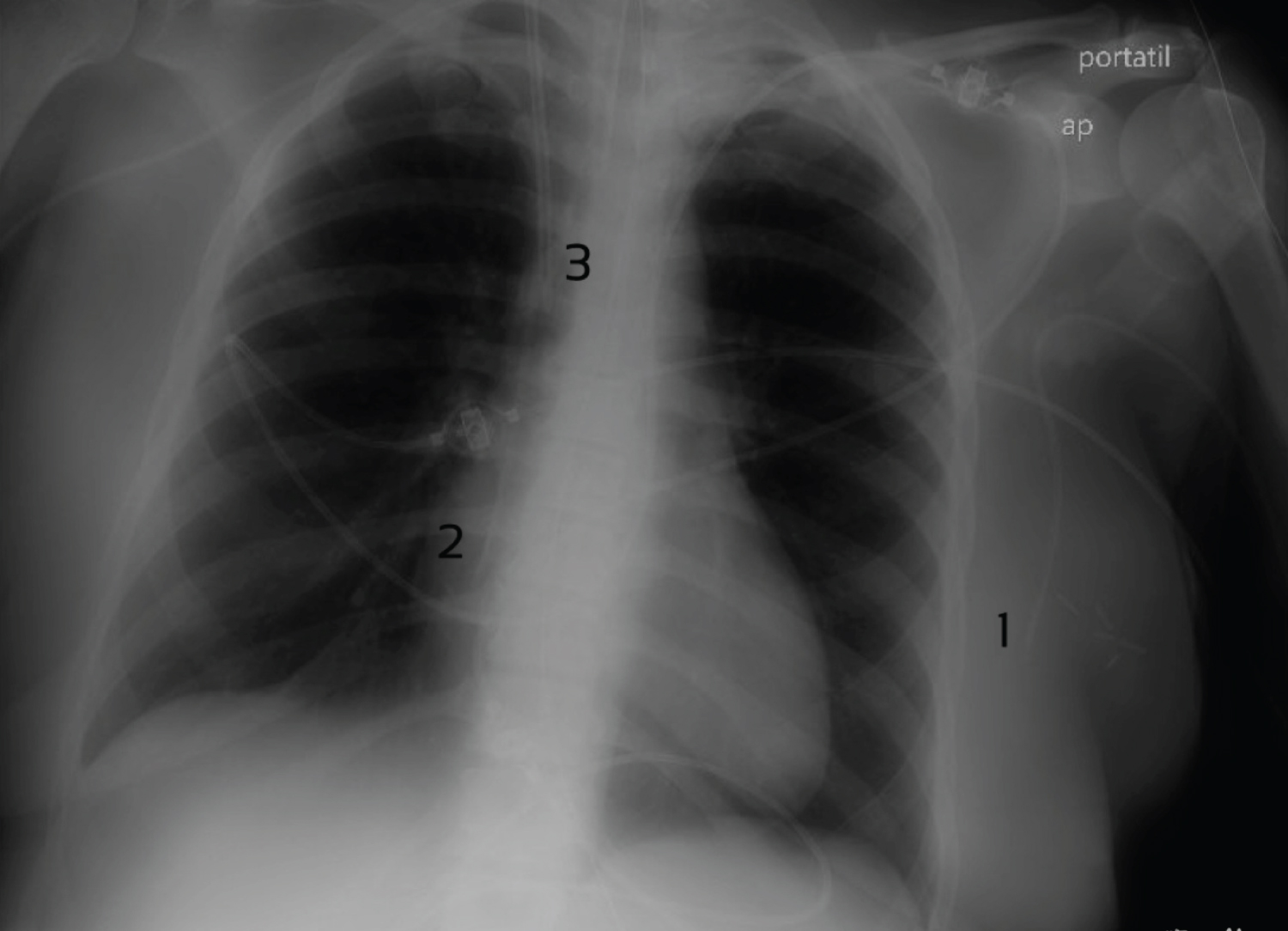
We present here an original image (Figure 1) of a portable chest radiograph where a PICC line inserted into the left upper arm can be identified as presenting its distal tip in the ipsilateral thoracodorsal vein (Figure 1, mark #1). It was placed in an obese, 45-year-old woman patient, suffering from cervical spinal cord injury and hospitalized in the Postoperative Intensive Care Unit after cervical arthrodesis. The aim was to replace the contralateral PICC, which was also malpositioned with its tip located in the right atrium (Figure 1, mark #2). These PICC lines were inserted through each basilic vein under echographic guidance. Both of them were later withdrawn without any complications and a new vascular access was achieved. Another intravascular device can be identified in the radiograph: A dialysis catheter is placed through the right internal jugular vein with its tip lying in the superior vena cava (Figure 1, mark #3).
If the tip of a central venous catheter is inserted into the heart chambers, there is a risk of arrhythmia, cardiac tamponade and heart valve damage. If the catheter tip is placed in a peripheral vein, phlebitis, vascular damage, extravasation and thrombosis may develop. For this reason, checking the position of the distal tip of PICC lines is mandatory. If malpositioned, the catheter should be repositioned or replaced [1,2]. Catheterization of the thoracodorsal vein during PICC placement is very unusual. Special precautions should be taken when placing a central catheter in patients with previous head and neck reconstruction surgery with pedicled latissimus dorsi flaps, as the lesion or thrombosis of the thoracodorsal vein or subclavian vein (to which the thoracodorsal vein is tributary) could trigger a flap failure [4].
PICC line placement should be always checked. Some unusual PICC tip placements should be adequately identified and corrected if necessary to avoid complications.
References
- Song L, Li H (2013) Malposition of peripherally inserted central catheter: Experience from 3,012 patients with cáncer. Exp Ther Med 6: 891-893.
- Kwon S, Son SM, Lee SH, et al. (2020) Outcomes of bedside peripherally inserted central catheter placement: A retrospective study at a single institution. Acute Crit Care 35: 31-37.
- Ng KS, Teh BT, Siew EP, et al. (1996) Malposition of a long central venous catheter in the right inferior thyroid vein--a case report. Singapore Med J 37: 556-558.
- Efanov JI, Bouhadana G, Danino MA (2019) Peripherally inserted central catheters as potential risk factors for failure in pedicled latissimus dorsi flaps. Plast Reconstr Surg Glob Open 7: e2236.
Corresponding Author
Enrique Sepúlveda Haro, Anesthesiology and Critical Care, Virgen de la Victoria University Hospital, Avenida Jorge Luis Borges, 51. 2℃, ZC: 29010, Málaga, Spain.
Copyright
© 2022 Sepúlveda HE, et al. This is an open-access article distributed under the terms of the Creative Commons Attribution License, which permits unrestricted use, distribution, and reproduction in any medium, provided the original author and source are credited.


