Death following a Massive Subcutaneous Emphysema
Abstract
Subcutaneous emphysema is a relatively common finding in clinical practice. It rarely leads to fatalities. The few reported deaths in the literature have all been associated with pneumothorax. We present the case of a 61-year male who developed a fatal massive subcutaneous emphysema on a background of end stage Chronic Obstructive Pulmonary Disease with Type 2 respiratory failure. This case highlights the fact that subcutaneous emphysema can be fatal even in the absence of a pneumothorax.
Introduction
Subcutaneous emphysema is typically a common, benign, and self-limiting complication of thoracic procedures. It does not usually require active management. Massive subcutaneous emphysema on the other hand usually requires active management to limit discomfort and minimize risk of respiratory compromise; nevertheless, it very rarely leads to death. We present the case study of a 61-year-old male who had a fatal outcome following a massive subcutaneous emphysema.
Case
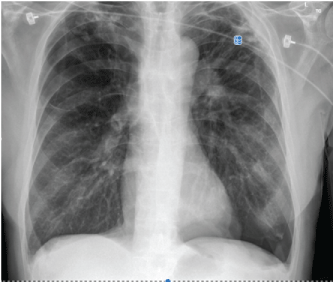
A 61-year-old male with known history of end stage chronic obstructive pulmonary disease, congestive heart failure and anxiety presented to the emergency department with acute onset respiratory distress and confusion. His GCS was 8, his respiratory rate was 40, and oxygen saturations were 70% on room air. His arterial blood gases revealed an acidemia with type 2 respiratory failure. After failing a trial of BiPAP, he was intubated via rapid sequence induction, and transferred to the intensive care unit where a large left sided pneumothorax was diagnosed (Figure 1).
Immediate needle decompression was performed followed by the placement of a pigtail intercostal catheter (ICC) connected to an underwater seal drain. The ICC was by bubbling and swinging, suggesting adequate placement of the drain. An ECG showed no ischemic changes, but biochemical assays revealed elevated levels of troponins, consistent with an acute coronary syndrome. He improved overnight and was extubated to BIPAP the following morning. He appeared to improve clinically over the next few days requiring only intermittent positive ventilator support (BiPAP). His chest x rays confirmed lung reinflation.
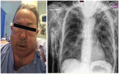
With continued improvement, he commenced self-attempts at mobilizing with his chest drain in-situ. Following one such attempt, he developed sudden onset respiratory distress. Further, he was noted to have developed extensive swelling in his chest, hands, face and bilateral palpebral fissures. He was unable to open his eyes and mouth. The clinical features were consistent with a massive subcutaneous emphysema, which was further confirmed on chest x-ray. Figure 2 shows a picture of the patient at the time of deterioration (left), as well as a picture of the chest x ray with massive subcutaneous emphysema (right).
All indications suggested that the subcutaneous emphysema may have developed due to dislodgment of the chest drain, most likely following the patient's attempts at self-mobilization. Further, exploration of the chest drain showed that it was also clogged with fibrin clots. Immediate attempts were made to aspirate the fibrin clots, and the patient was immediately commenced on positive pressure ventilation, while a formal new chest drain was inserted. Low pressure suction was applied to the drain and despite continuous bubbling, the massive subcutaneous emphysema prevented the patient from opening his eyes and mouth.
There was no improvement over the next few days. The patient consistently refused to lie in bed, as he felt much more comfortable spending the whole day and night in a sleeping chair in the tripod position. He complained of inability to breath and to open his mouth and eyes. The inability to open his mouth did not allow for intubation or use of airway adjuncts. He was hence kept on BiPAP.
Both the patient and his family declined further aggressive management. He was hence palliated and passed away a few hours later.
Discussion
The development of subcutaneous emphysema in our patient was most likely a direct consequence of his chest drain dislodgement while mobilizing. Subcutaneous emphysema is typically a benign, self-limiting condition, which occurs when penetrating or blunt force trauma causes air to track from the interstitial tissues surrounding the pulmonary vasculature and into the hilum, thereby leading to development of pneumomediastinum. From here, the air may then track into the soft tissues of the neck, face and chest wall, leading to clinical complications such as significant swelling, dyspnea, dysphagia and dysphonia [1]. Emphysema can arise due to any of three situations, namely: (a) Gaseous forming microorganisms during an infection, (b) Direct introduction of air into the soft tissues following traumatic disruption of cutaneous or mucosal barriers, and (c) Spontaneously, when there is a sufficient pressure difference between the air-filled alveolus and the surrounding interstitial space which leads to alveolar rupture [2].
The soft tissues of the neck are divided into three compartments by the deep cervical fascia. The visceral space invests the trachea and esophagus. This creates a direct communication between the neck and the chest. Inferiorly, the visceral space communicates with the retroperitoneal soft tissue space by following the esophagus through the diaphragmatic hiatus [2].
There is hence a continuum connecting the neck, chest and abdomen [2].
A review of the literature suggests that death from subcutaneous emphysema is extremely rare, with the few reported deaths occurring in the context of pneumothorax. In particular Johnson, et al. [3] reviewed several cases of subcutaneous emphysema, including the management techniques used, and the subsequent outcomes. No deaths were reported following management of these patients.
Death from subcutaneous emphysema in the presence of pneumothorax has been attributed to 2 possible mechanisms. Air may compress the great vessels and the airway in the hypopharynx, leading to hemodynamic instability, and subsequently death. Alternatively, if the subcutaneous emphysema occurs in the context of a pneumothorax, the subsequent development of a tension in the pneumothorax may lead to decreased cardiac output and eventually death [4].
Several studies have explored optimal modalities for treatment of extensive subcutaneous emphysema [3]. The most common approaches include infraclavicular incisions, subcutaneous drainage, and suction on in-situ chest drains [3]. High flow oxygen therapy may also be employed to facilitate resorption of nitrogen from distended tissues [5]. In our case, intubation was not possible because the patient could not open his mouth and had developed extensive neck swelling. The combination of ongoing respiratory failure, hemodynamic instabilities, and anxiety led to continuous distress for the patient, hence the decision was made to commence him on positive pressure ventilation and place a formal chest drain. He unfortunately did not improve in the days following the onset of the massive emphysema. Both him and his family refused to have further invasive management of his respiratory failure and massive subcutaneous emphysema.
Conclusion
Subcutaneous emphysema is a relative common finding in clinical practice, and rarely leads to fatality. The very few reported subcutaneous emphysema related deaths in the literature have all been associated with pneumothorax. This case report highlights the fact that subcutaneous emphysema can be fatal, even in the absence of a pneumothorax. Clinical vigilance is warranted.
Author Contributions
The manuscript has been read and approved by all the authors, that the requirements for authorship as stated earlier in this document have been met. Each author believes that the manuscript represents honest work.
References
- O'Reilly P, Chen HK, Wiseman R (2013) Management of extensive subcutaneous emphysema with a subcutaneous drain. Respirol Case Rep 1: 28-30.
- Maunder RJ, Pierson DJ, Hudson LD (1984) Subcutaneous and mediastinal emphysema. Pathophysiology, diagnosis and management. Arch Intern Med 144: 1447-1453.
- Johnson CH, Lang SA, Bilal H, et al. (2014) In patients with extensive subcutaneous emphysema, which technique achieves maximal clinical resolution: Infraclavicular incisions, subcutaneous drain insertion or suction on in situ chest drains? Interactive Cardiovasc Thorac Surg 18: 825-829.
- Peatfield RC, Edwards PR, Johnson NM (1979) Two unexpected deaths from pneumothorax. Lancet 1: 356-358.
- O'Neill TJ, Johnson MC, Edwards DA, et al. (1979) Ventilation with 100 percent oxygen for life-threatening mediastinal and subcutaneous emphysema. Chest 76: 492-493.
Corresponding Author
Dr. Avisham Ramphul, Division of Surgery, John Hunter Hospital, Newcastle, NSW, Australia.
Copyright
© 2018 Ramphul A, et al. This is an open-access article distributed under the terms of the Creative Commons Attribution License, which permits unrestricted use, distribution, and reproduction in any medium, provided the original author and source are credited.