The Unusual Presentation of Acute Appendicitis in Pregnancy
Abstract
Acute appendicitis is the most common non-obstetric surgical emergency during pregnancy, and can be difficult to identify due to its unusual and inconsistent signs and symptoms.
We present a case of a young gravid lady with an inconsistent septic picture and abdominal signs. Computed Tomography (CT) imaging was used to identify the cause as acute appendicitis.
In this case study, we discuss the following:
1. The use of high-risk diagnostic imaging in a pregnant abdomen.
2. Different sites of incisions as described in literature and their justification.
3. The use and safety of laparoscopic and 'open' approach in a pregnant patient.
Introduction
Acute appendicitis is the most common non-obstetric surgical emergency during pregnancy, resulting in devastating consequences on both the mother and the fetus [1,2]. The incidence of appendicitis in pregnancy is similar to that of the general population. However, the clinical presentation and diagnosis of a pregnant patient with acute appendicitis can be difficult, and may present without the common signs and symptoms that the general population present with [3].
Case
A 21-year-old women (G2P1 - Gravid a 2 Parity 1), twenty-seven weeks gestational age was admitted through her midwife with a one day history of unlocalised left sided abdominal pain, emesis and a septic picture. Temperature 38 ℃, Heart rate 120 bpm, Blood pressure 78/42 mmgH.
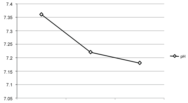
Initial blood results revealed raised white cell count of 16.3 × 10⁹ per Litre (L) and a raised C-reactive protein levels. Arterial blood gas showed mild metabolic acidosis with respiratory compensation; electrolytes and liver function values were within normal limits. Cardiotocography showed no contractions.
Due to this unusual presentation and left localising signs, the initial diagnosis was renal/pelvic pathology. Ultrasound examination showed evidence of slight left hydronephrosis, which did not account for the patients clinical and biochemical profile.
Due to the increased inflammatory markers and abnormal bloods gases (Graph 1), the patient was admitted to the high dependence unit for close observations. Due to the unstable nature of the patient's condition, an abdominal CT scan was requested to help identify the source of sepsis.
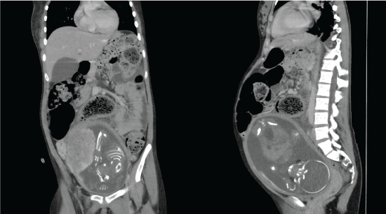
Abominal CT scan demonstrated a fluid filled appendix with wall enhancement (Figure 1) as well as free fluid in the abdomen. Given the suspicion of acute appendicitis from the scan, the patient was listed for an emergency open appendectomy.
The patient was put under general anesthesia; consultant obstetrician/gynecologist and the surgical registrar were present.
The incision was made through a higher transverse lanz superior to McBurney’s point; an inflamed appendix, free pus, dilated cecum and ileum were observed in the abdomen. No operative complications occurred.
The patient recovered well, and was discharged after a total of 7 days post-operative.
Discussion
Appendicitis is clinical diagnosis, which may not require imaging if there is high clinical suspicion as it may delay necessary surgical intervention.
Ultrasound scanning is considered to be the first line of imaging [4], followed by CT scans.
CT scans have improved the efficiency of diagnosing acute appendicitis in the general population compared to the use of ultrasound imaging [5,6].
CT scans have improved the efficiency acute appendicitis diagnosis in the general population in contrast to ultrasound scanning [5,6].
Efficiency of diagnostic CT imaging (Table 1) has influenced the certainty when formulating a definite management plan for patients presenting with a non-traumatic acute abdomen [7].
In pregnancy, the use of CT imaging should be very carefully considered, due to the risk of radiation exposure to the fetus [2]. The risk of radiation from CT scans ranges between 1 to 5 rads, which is below the radiation exposure that may cause teratogenic effects (5-15 rads) according to a study by Chen, et al. [4].
The fetus is potentially it’s most vulnerable at 16 weeks of gestation. A single dose of CT scan radiation may stimulate a spontaneous abortion.
Cumulative radiation exposure also increases the risk of severe mental retardation (40%) and tumor induction (15% life span risk) [2,8].
Magnetic Resonance Imaging (MRI), is an acceptable substitution for CT scanning in pregnancy. A study by Cobben, et al. [9] stated that MRI scans for suspected appendicitis can be achieved in room time of less than 20 minutes without the administration of gadolinium.
Therefore, MRI scan can be used effectively, without the risks of radiation exposure experienced in CT scans [9]. Nevertheless, MRI should be avoided in the first trimester, as the fetus is highly vulnerable to any unknown bio-effects of MRI [9,10].
In our case, the use of MRI scan over a CT was not practical due to the deteriorating condition the patient and the time constraint.
There are conflicting views in the current literature in regards to the anatomical positioning of the appendix during pregnancy. Counter clockwise rotational displacement of the appendix and cecum in gravid patients have been observed by Baer, et al. [11] using barium enemas, however, results have not been replicated due to the risk of radiation [11-13]. Hodjati, et al. [12] disagrees with the degree of displacement with the gestational age observed by Baer, et al. [11] but does report a degree of displacement of 3-4 cm from McBurney’s point [12].
The degree of anatomical displacement is the crucial factor that influences the site of optimal incision needed for appendectomy. Popkin, et al. [13] demonstrates that it is easier to locate the appendix in a pregnant abdomen through an incision over McBurney’s point (94%) compared to incisions made superior to the McBurney’s point (80%) regardless of the gestational age [13]. However, the study by Popkin, et al. [13] has been limited due to the variation of skills amongst surgeons and the small number of cases observed. In our case, the appendix was located caudal to the incision, and superior to McBurney’s point, which suggested a degree of upwards displacement of the appendix caused by the gravid uterus.
In our case study and in a case study done by Donkervoort, et al. [14] the appendix was displaced from its typical anatomical position due to a gravid uterus. This would not have been present at a McBurney’s incision and will result in the prolongation of surgery and time to find the displaced appendix, resulting in a larger scar.
The guidelines published by The Society of American Gastrointestinal and Endoscopic Surgeons for surgical problems in pregnancy state that the laparoscopic approach is the preferred method in pregnant patients with suspected appendicitis. These guidelines have been advised regardless of gestational age, giving the same benefits as non-pregnant patients. These results in decreased length of hospital admission and less postoperative pain [15-17]. Even though the laparoscopic approach in pregnancy is associated with a low rate of intraoperative complications in all three trimesters, it has been associated with a higher (6%) rate of fetal loss compared to open appendectomy [14,16]. The trocars insertion points are shifted upwards towards the subcoastal regions depending on the gestational age to avoid uterine injury [12,14,15].
The surgical registrar had to take the patient into theatre for an emergency open appendectomy, out of hours, using a Lanz incision superior to McBurney’s point. An ‘open’ appendectomy approach was chosen over laparoscopic approach due to limited surgical experience.
Conclusion
Appendicitis is based on clinical diagnosis and imaging is not always required. Ultrasound imaging should always be considered as a first line, in the case of inconclusive results, additional imaging should be considered.
A single CT has been shown to pose minimal risk to the fetus, but the radiation risk should be weighed against the benefit and discussed with the mother [2]. MRI scans are an alternative to CT imaging, and adequate images can be achieved within 20 minutes. MRI has been reported to have very high accuracy in detecting a positive finding in early pregnancy. Nevertheless, accuracy of MRI is decreased in late pregnancy. The use of MRI should be limited in the first trimester due to assumed risk [9]. The use of higher risk imaging such as CT or MRI should be discussed in a multidisciplinary manner with obstetrics and gynecology, paediatrics, radiology, surgical team and the parents.
Laparoscopic appendectomies in pregnant women have a low rate of complication in the hands of a skilled surgeon. Surgeons’ operate at their preference, and use techniques based on their skills, competence and comfort. The options for appendectomies approach in pregnancy are: ‘open’ using a Lanz incision, a midline laparotomy incision or a laparoscopy approach.
Further studies are required to establish the best anatomical landmark of incision on pregnant patients with appendicitis. The evidence of using a McBurney’s incision as suggested by Popkin, et al. [13] alternatively to an incision superior to McBurney’s point [11] is very limited.
Conflict of Interest
No conflict of interest or funding declared.
References
- Sakhri J (2001) Acute appendicitis during pregnancy. Tunis Med 79: 521-525.
- Murariu D, Tatsuno B, Hirai CA, et al. (2011) Case report and management of suspected acute appendicitis in pregnancy. Hawaii Med J 70: 30-32.
- Retzke U, Graf H, Schmidt M (1998) Appendicitis in pregnancy. Zentralbl Chir 4: 61-65.
- Chen MM, Coakley FV, Kaimal A, et al. (2008) Guidelines for computed tomography and magnetic resonance imaging use during pregnancy and lactation. Obstet Gynecol 112: 333-340.
- Balthazar EJ, Birnbaum BA, Yee J, et al. (1994) Acute appendicitis: CT and US correlation in 100 patients. Radiology 190: 31-35.
- Malone AJ Jr, Wolf CR, Malmed AS, et al. (1993) Diagnosis of acute appendicitis: value of unenhanced CT. AJR Am J Roentgenol 160: 763-766.
- Abujudeh HH, Kaewlai R, McMahon PM, et al. (2011) Abdominopelvic CT increases diagnostic certainty and guides management decisions: a prospective investigation of 584 patients in a large academic medical center. AJR Am J Roentgenol 196: 238-243.
- Kal HB, Struikmans H (2002) Pregnancy and medical irradiation; summary and conclusions from the International Commission on Radiological Protection, Publication 84. Ned Tijdschr Geneeskd 146: 299-303.
- Cobben LP, Groot I, Haans L, et al. (2004) MRI for clinically suspected appendicitis during pregnancy. AJR Am J Roentgenol 183: 671-675.
- Shellock FG, Kanal E (1991) Policies, guidelines, and recommendations for MR imaging safety and patient management. SMRI Safety Committee. J Magn Reson Imaging 1: 97-101.
- Baer JL, Reis RA, Arens RA (1932) Appendicitis in pregnancy with changes in position and axis of the normal appendix in pregnancy. JAMA 98: 1359-1364.
- Hodjati H, Kazerooni T (2003) Location of the appendix in the gravid patient: a re-evaluation of the established concept. Int J Gynaecol Obstet 81: 245-247.
- Popkin CA, Lopez PP, Cohn SM, et al. (2002) The incision of choice for pregnant women with appendicitis is through McBurney's point. Am J Surg 183: 20-22.
- Donkervoort SC, Boerma D (2011) Suspicion of acute appendicitis in the third trimester of pregnancy: pros and cons of a laparoscopic procedure. JSLS 15: 379-383.
- (2011) Guidelines for Diagnosis, Treatment, and Use of Laparoscopy for Surgical Problems during Pregnancy. SAGES Society of American Gastrointestinal and Endoscopic Surgeons.
- Machado NO, Grant CS (2009) Laparoscopic Appendicectomy in all Trimesters of Pregnancy. JSLS 13: 384-390.
- Lemieux P, Rheaume P, Levesque I, et al. (2009) Laparoscopic appendectomy in pregnant patients: a review of 45 cases. Surg Endosc 23: 1701-1705.
Corresponding Author
A Arnaout, Foundation Trainee, Department of General Surgery, Warrington and Halton NHS Foundation Trust Lovely Ln, University of Buckingham Medical School, Warrington WA5 1QG, Buckingham, UK.
Copyright
© 2017 Arnaout A, et al. This is an open-access article distributed under the terms of the Creative Commons Attribution License, which permits unrestricted use, distribution, and reproduction in any medium, provided the original author and source are credited.