Acute Flank Pain in the Emergency Room: Rare Case and Review of the Literature
Abstract
Wunderlich syndrome is defined as spontaneous renal hemorrhage. Generally, the kidney mass may be due to secondary causes such as vascular pathologies.Wunderlich syndrome is rarely idiopathic. Wunderlich syndrome is a classical clinical symptom of Lenk's triad, characterized by flank pain, mass, and hypotension. Wunderlich syndrome can be life threatening due to gross hemorrhage. Our patient applied with the complaint of abdominal pain. A hemorrhage area was detected in the area corresponding to the kidney location in abdominal ultrasonography upon the presence of sensitivity in lower right scale detected in the examination. Hemorrhage area was detected in abdominal contrast-enhanced tomography. Our patient was discharged without any complications after following up conservatively. Wunderlich syndrome, which can rarely be diagnosed in emergency departments, should be considered in the differential diagnosis of emergency physicians.
Introduction
Spontaneous nontraumatic idiopathic renal hemorrhage is known as Wunderlich Syndrome (WS) [1]. Patients with WS may go to emergencys ervices in a wide range of conditions, from mild to severe, life-threatening clinical conditions. The severity of the disease varies depending on the extent of the bleeding into the retroperitoneal area.
Renal hemorrhage, which may be secondary to underlying etiologic factors, is a well-known clinical entity. However, information on idiopathic and spontaneous renal hemorrhages is very little in the literature [2]. Here, we aimed to present a case of spontaneous renal hemorrhage in a patient whose vital signs were completely normal without any risk factors.
Case
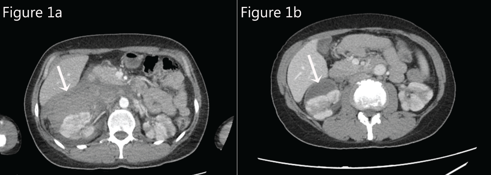
A 37-year-old female patient was admitted to our emergency department with the complaint of flank pain starting suddenly the day before. She stated that after the pain started in the right flank area, it gradually widened, spreading to her back and in to her stomach. Her pain was insistent, and its severity gradually increased. She did not mention any urinary system complaints, including hematuria. She stated that she never had similar painful and localized pain like this before. She had no trauma story. There was no history of chronic illness in her medical history. The vital signs of the patient were in normal range. (TA: 122/71 mmHg, SpO2: 98%, pulse: 77/ min, temperature: 37 ℃). Physical examination revealed right costovertebral angle sensitivity. With palpation, there was no defense or rebound while the abdomen was sensitive on the right upper and lowerside. It was in evidence that the hemoglobin values were lower in the blood parameters of the patient (hemoglobin: 8.5 g/dl), it was observed as RBC: 27.00/HPF in the urine test, all other laboratory values including the white blood cell were in the normal range. Abdominal and urinary system USG was requested in order to eliminate the causes of abdominal pain. USG was reported as an area compatible with hemorrhage in the right kidney. Similar retroperitoneal hemorrhage was observed in the right kidney in the requested abdominal tomography in order to confirm the diagnosis (Figure 1a). At the CT scan showed that normal renal parenchyma. No performed renal angiography. According to USG, BT scan and lab findings there was no alternative diagnosis. The patient who was admitted to the urology clinic for consultation in terms of spontaneous renal hematoma was admitted to the same clinic. Symptomatic treatment was applied here. No histological examination was performed because the patient was symptomatic manegement. The clinical condition improved and she was discharged on the 5th day of the hospitalization.
It was seen that the bleeding was organized and the area was reduced in the abdominal tomography taken in the patient who was called to the control 2 months after the discharge (Figure 1b).
Discussion
A renal hematoma can occur secondary to many causes such as renal neoplasms, vascular kidney diseases, renal cystic diseases, renal stones and coagulation disorders. A spontaneous renal hematoma (Wunderlich syndrome) that is not related to any cause is a very rare clinical condition [2]. Classically, Wunderlich's syndrome is characterized by the presence of the Lenk's triad including acute flank pain and/or back pain, mass and hypotension involved, tachycardia, anemia [3]. However, it is very rare that these three symptoms appear at the same time. Patients usually apply with symptoms such as abdominal pain (67%), hematuria (40%) and hypovolemic shock (26.5%) [1]. Other common symptoms include nausea, vomiting, low-grade fever, and anemia [4]. In our case, surprisingly, vital findings were in a perfectly normal range and our patient had no complaints other than abdominal and flank pain. Further more, the absence of a risk factor for renal hemorrhage in our patients was a diagnostic challenge. In the case reported by Medda, et al. hypovolemic shock due to renal hematoma developed and hemoglobin values of the patient decreased from 13.5 g/dL to 8.5 g/dL [5]. Similarly, although our patient had a hemoglobin value of 8.5, our patient did not develop any clinical evidence of hypovolemic shock. The fact that the age and body mass indexes of two patients are different from each other may be the reason.
The most common etiologic cause of renal hematoma is neoplasia. The underlying cause in 60-65% of all cases is renal neoplasms. Angiomyolipoma is the most common benign neoplasm responsible for renal hematoma. Renal cell carcinoma is the most common malignant neoplasm. 6.7% of all kidney hemorrhagic cases are idiopathic [6]. Baishya, et al. suggested in their case that uncontrolled hypertension may be a risk factor for the development of a spontaneous renal hematoma [4]. As in our case, the spontaneous renal hematoma is rarely seen in which there is no known risk factor.
Spontaneous renal hematomas are usually detected by imaging when investigating other causes of lumbar pain and hypotension. In addition to laboratory, radiology is important for diagnosis. Ultrasonography, computed tomography, magnetic resonance imaging and angiography are used. Although the conservative approach is advocated in the treatment [4], capsulotomy, percutaneous drainage, nephrectomy and radiologic embolization are applied according to the patient's clinic. In our case, USG was performed and the renal hematoma was detected while investigating the potential causes of abdominal pain. The diagnosis was clarified by abdominal CT for detection of bleeding center and differential diagnosis. Conservative treatment was planned on the basis of the stability of the patient's clinic in our case. After conservative treatment, the patient was discharged without any complications.
In conclusion, clinical suspicion is very important for the diagnosis of Wunderlich Syndrome. The low incidence of Wunderlich Syndrome may be due to the lack of clinical suspicion and the difficulty of diagnosis. The life-threatening Wunderlich Syndrome should be considered in the differential diagnosis of patients applied to the emergency service with flank pain.
References
- Zhang JQ, Fielding JR, Zou KH (2002) Etiology of spontaneous perirenal hemorrhage: a meta-analysis. J Urol 167: 1593-1596.
- Katabathina VS, Katre R, Prasad SR, et al. (2011) Wunderlich syndrome: cross-sectional imaging review. J Comput Assist Tomogr 35: 425-433.
- Mao Y, De Oliveira IS, Hedgire S, et al. (2017) Aetiology, imaging features, and evolution of spontaneous perirenal haemorrhage. Clin Radiol 72: 175.e19-175.e26.
- Baishya RK, Dhawan DR, Sabnis RB, et al. (2011) Spontaneous subcapsular renal hematoma: A case report and review of literature. Urol Ann 3: 44-46.
- Medda M, Picozzi SC, Bozzini G, et al. (2009) Wunderlich's syndrome and hemorrhagic shock. J Emerg Trauma Shock 2: 203-205.
- Greco M, Butticè S, Benedetto F, et al. (2016) Spontaneous Subcapsular Renal Hematoma: Strange Case in an Anticoagulated Patient with HWMH after Aortic and Iliac Endovascular Stenting Procedure. Case Rep Urol 2016: 2573476.
Corresponding Author
Abdullah Osman Kocak, MD, Emergency Department of Medical Faculty, Ataturk University, 25240, Erzurum, Turkey, Tel: +90-442-3448401, Fax: +90-442-3443133.
Copyright
© 2017 Kocak AO, et al. This is an open-access article distributed under the terms of the Creative Commons Attribution License, which permits unrestricted use, distribution, and reproduction in any medium, provided the original author and source are credited.