Utility of P16 and Ki-67 Expression in Cervical Biopsy with Cervical Intraepithelial Neoplasia 2 to Predict Diagnosis in Subsequent Cervical Excision Specimen
Abstract
Treatment guidelines differ for young women with Cervical Intraepithelial Neoplasia (CIN) 2 lesions on biopsy depending on their desire for future pregnancy. Therefore, we investigated the utility of using P16 and Ki-67 immunohistochemical stains on cervical biopsies diagnosed with CIN 2 to determine the outcome on subsequent excisional specimens. The study included 22 patients, with a mean age of 34 years and an age range from 20 to 51 years. Positive staining for p16 was identified in 21/22 patients. The subsequent excisional pathology of these 21 patients with strong diffuse staining showed that 1 had invasive squamous cell carcinoma, 9 had CIN 3, 6 had CIN 2 or CIN 1-2, 2 had CIN 1, and 3 had no CIN lesion. The only patient with patchy p16 staining on cervical biopsy had CIN 3 on cone biopsy. The Ki-67 index was negative in 18/22 patients and positive in 4/22 patients. The Ki-67 index was negative in 5/5 patients with CIN 1 or no CIN lesion on subsequent excisional pathology and 13/17 patients with CIN 2 or CIN 3 on follow up excisional pathology. The 4 patients with a positive Ki-67 index all had CIN 2 or CIN 3 on follow up excisional pathology. Thus, we conclude that P16 and Ki-67 immunostaining was not found to be useful in triaging women with CIN 2.
Keywords
Cervical Intraepithelial Neoplasia 2 (CIN 2), Ki-67, P16, Human Papillomavirus (HPV), Cervix
Introduction
Human Papillomavirus (HPV) is a double stranded DNA virus with over 150 subtypes. Infection with some of these subtypes, most commonly HPV 16 and HPV 18, can lead to cancer in multiple body sites, including the cervix, vagina, vulva, penis, anus and oropharynx. The Centers for Disease Control and Prevention reported that HPV is the cause of over 90% of cervical cancers [1].
HPV infects skin and mucosal surfaces, and after infection it can cause squamous epithelium to undergo premalignant changes that can eventually lead to Squamous Cell Carcinoma (SCC). The progression from premalignant HPV driven lesions to SCC does not occur in all patients. Although a significant portion of CIN 2 cases progress to CIN 3, regression of CIN 2 is not uncommon. A study by Nasiell, et al. [2] reported that 54% of CIN 2 cases regressed, 16% persisted, and 30% cases progressed to CIN 3. As of yet, there is no definitive way to determine which CIN 2 lesions will progress and which will regress.
The Lower Anogenital Squamous Terminology (LAST) Standardization Project for HPV-Associated Lesions recommended that cervical HPV-associated squamous lesions be called Low-Grade Squamous Intraepithelial Lesion (LSIL) and High-Grade Squamous Intraepithelial Lesion (HSIL), and that these lesions may be further subclassified as CIN 1, CIN 2, or CIN 3 [3]. The American College of Obstetricians and Gynecologists and American Society for Colposcopy and Cervical Pathology guidelines recommend that young women with CIN 2 lesions on biopsy can be treated with either observation (if they desire future pregnancy) or excision/ablation of the transformation zone [4,5]. Thus, it will be of great clinical significance to look for biomarkers which can predict the clinical outcome and guide the management of CIN 2.
It has been previously proven that the use of the immunohistochemical stains p16 and Ki-67 can improve the diagnostic accuracy of CIN 2 and CIN 3 [6-10]. In addition, in the LAST Standardization Project for HPV-Associated Lesions it is recommended that p16 immunohistochemistry should be used when a cervical biopsy is histomorphologically a CIN 2 in order to help determine if the lesion should be considered a low or a high grade lesion [3]. Therefore, we postulated that p16 and Ki-67 stains may have predictive value in patients with cervical biopsies with CIN 2.
Materials and Methods
The NYU Hospital laboratory information system was searched for cervical biopsy cases with a histological diagnosis of CIN 2 that were accessioned between 2004 and 2010; these dates were randomly chosen in order to obtain enough cases. Cases were included in this study if there was a subsequent excisional specimen (Loop Electrosurgical Excision Procedure [LEEP] or Cone Biopsy), and cases were excluded from the study if the dysplastic area was no longer present in deeper levels obtained for immunohistochemical stains.
Immunohistochemical stains for p16 and Ki-67 were performed on adjacent 4-µm sections of formalin-fixed, paraffin-embedded. The primary antibodies were unconjugated monoclonal mouse anti-human p16 INK4a (Ventana Medical Systems, Tucson, AZ) and unconjugated rabbit anti-human Ki67 clone SP7 (Lab Vision, Waltham, MA).
The p16 immunohistochemical stain was graded as follows: The p16 stain was considered positive if there was diffuse and strong staining (continuous strong nuclear or nuclear plus cytoplasmic staining of the basal and parabasal cell layers) and all other patterns were considered negative [11].
A previous study showed that a combination of the quantity and distribution of Ki-67 positive cells within the squamous epithelium correlated with whether the cervical biopsy was LSIL or HSIL. The Ki-67 immunohistochemical stain was graded as follows: The Ki-67 index was considered positive if the squamous epithelium had at least a 30% index in either the basal layer or the upper 1/3 of the squamous epithelium (associated with HSIL), and all other staining patterns were considered to be negative (associated with LSIL) [12].
Results
A total of 22 patients qualified for the study, with a mean age of 34 years and an age range from 20 to 51 years. The average time interval between the initial cervical biopsy and the subsequent excisional procedure was approximately 2 months and the range was from 1 to 4 months. The follow up excisional pathology demonstrated at least CIN 2 in 17/22 patients, and CIN 1 or no CIN lesion in 5/22 patients.
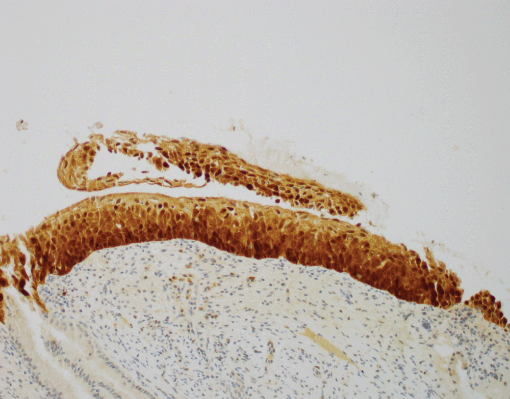
Positive staining for p16 was identified in 21/22 patients (Figure 1). At least 50% of the height of the epithelium was stained in 21/21 cases and the entire epithelium was stained in 15/21 cases. The subsequent excisional pathology of these 21 patients with strong diffuse staining showed that 1 had invasive squamous cell carcinoma, 9 had CIN 3, 6 had CIN 2 or CIN 1-2, 2 had CIN 1, and 3 had no CIN lesion. The only patient with negative p16 staining on cervical biopsy had CIN 3 on subsequent excisional pathology (Table 1).
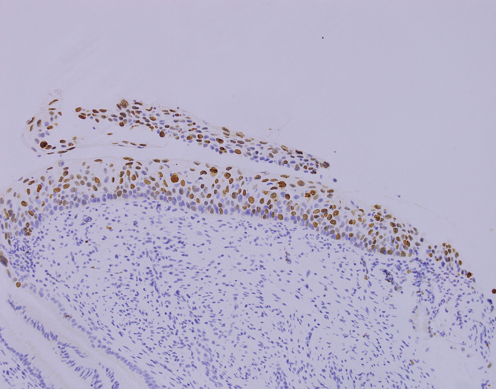
The Ki-67 index was negative in 18/22 patients and positive in 4/22 patients (Figure 2). The Ki-67 index was negative in 5/5 patients with CIN 1 or no CIN lesion on subsequent excisional pathology and 13/17 patients with CIN 2 or CIN 3 on follow up excisional pathology. The 4 patients with a positive Ki-67 index all had CIN 2 or CIN 3 on follow up excisional pathology (Table 1). The staining pattern in the 4 positive patients are as follows: 2 had staining only in the upper 1/3 of the squamous epithelium, 1 had staining only in the basal layer of the squamous epithelium, and 1 had staining in both the upper 1/3 and the basal layer of the squamous epithelium. The Ki-67 indices were all around 30-40%, except 1 case that had a Ki-67 index of 70%.
Discussion
Our study found that p16 immunohistochemistry is unable to predict subsequent excisional pathology findings in patients with cervical biopsies with CIN 2. A positive staining pattern for p16 was seen in almost all (21/22) biopsies with CIN 2. Only one biopsy lacked such expression, and the subsequent excisional pathology was diagnosed as CIN 3. Thus, in our study, nearly all of the biopsies stained positive with P16 and thus it is not a useful test.
In our study 4/4 patients with positive Ki-67 indices had at least CIN 2 on follow up excisional pathology; however, only a small number of cases had a positive Ki-67 index, and thus further study is needed to determine the significance of this finding. A negative Ki-67 index is not specific and does not indicate a greater likelihood of regression or progression on subsequent excisional pathology. In our study 5/5 patients with CIN 1 or no CIN lesion on follow up excisional pathology had negative Ki-67 indices, but 13 patients with negative Ki-67 indices had at least CIN 2 on follow up excisional pathology.
Previous studies have found that both Ki-67 and p16 have predictive value for cervical biopsies.
A prior study by Kruse, et al. [13] showed that quantitative Ki-67 index measurements were better than the histologic CIN classification system at predicting subsequent CIN 3 on follow up procedures when the initial biopsy was diagnosed as CIN 1 (25 cases) or CIN 2 (65 cases). The study used the QPRODIT (version 6.1) interactive image analysis system to evaluate the Ki67 index in the middle third layer of the squamous epithelium. The study found that when using the quantitative Ki67 model 0/40 "Ki67-model low-risk" patients progressed to CIN 3, in contrast to 15/50 (30%) "Ki67-model high-risk" patients progressed to CIN 3 (p < 0.001). The study also found that subjective estimations of Ki67 were also prognostic, but not statistical significance (p = 0.06)
A study by Miralpeix, et al. [14], that included 123 patients, showed that CIN 3 in a LEEP can be predicted by a Ki-67 index of > 50% in a previous cervical biopsy diagnosed as CIN 2 (p = 0.043). In addition, this study also found that evaluation of p16 and Ki67 by thirds of the epithelial thickness in CIN 2 cervical biopsies did not correlate significantly with subsequent LEEP results.
In contrast to the result of our study, a prior study by Omori, et al. [15] showed that evaluating the immunohistochemical stain p16 and high-risk HPV In-Situ Hybridization (ISH) expression patterns in cases of CIN 2 may be useful in predicting progression. This study included 52 cases of CIN 2 diagnosed via cervical biopsy and found that stronger p16 immunohistochemical expression and a higher frequency of high-risk HPV ISH punctate nuclear signal expression significantly correlated with progression of CIN 2 to CIN 3.
The different studies may have different results because of different immunohistochemical staining procedures, different immunohistochemical antibody clones, inter-pathologist variability in diagnosing CIN 2, and the differing abilities of gynecologists to obtain cervical biopsies from the location of the lesion [16-18]. Standardization of these factors may be necessary before P16 and Ki67 can be applied as a predictive tool across many laboratories.
This study attempted to triage women with CIN 2 into one group that needs an excisional procedure and another group that can be treated with watchful waiting. P16 and Ki-67 immunostaining was not found to be useful in triaging women with CIN 2.
References
- https://www.cdc.gov/cancer/hpv/statistics/index.htm
- Nasiell K, Nasiell M, Vaclavinkova V (1983) Behavior of moderate cervical dysplasia during long-term follow-up. Obstet Gynecol 61: 609-614.
- Darragh TM, Colgan TJ, Cox JT, et al. (2012) The lower anogenital squamous terminology standardization project for HPV-associated lesions: Background and consensus recommendations from the college of american pathologists and the american society for colposcopy and cervical pathology. Arch Pathol Lab Med 136: 1266-1297.
- Massad LS, Einstein MH, Huh WK, et al. (2013) 2012 Updated consensus guidelines for the management of abnormal cervical cancer screening tests and cancer precursors. J Low Genit Tract Dis 17: S1-S27.
- American College of Obstetricians and Gynecologists (2013) Practice Bulletin No. 140: Management of abnormal cervical cancer Screening test Results and cervical cancer precursors. Obstet Gynecol 122: 1338-1367.
- Dray M, Russell P, Dalrymple C, et al. (2005) p16 (INK4a) as a complementary marker of high-grade intraepithelial lesions of the uterine cervix. I: Experience with squamous lesions in 189 consecutive cervical biopsies. Pathology 37: 112-124.
- Tsoumpou I, Arbyn M, Kyrgiou M, et al. (2009) p16(INK4a) immunostaining in cytological and histological specimens from the uterine cervix: A systematic review and meta-analysis. Cancer Treat Rev 35: 210-220.
- Gustinucci D, Passamonti B, Cesarini E, et al. (2012) Role of p16(INK4a) cytology testing as an adjunct to enhance the diagnostic specificity and accuracy in human papillomavirus-positive women within an organized cervical cancer screening program. Acta Cytol 56: 506-514.
- Galgano MT, Castle PE, Atkins KA, et al. (2010) Using biomarkers as objective standards in the diagnosis of cervical biopsies. Am J Surg Pathol 34: 1077-1087.
- Sari Aslani F, Safaei A, Pourjabali M, et al. (2013) Evaluation of Ki67, p16 and CK17 Markers in differentiating cervical intraepithelial neoplasia and benign lesions. Iran J Med Sci 38: 15-21.
- Bergeron C, Ordi J, Schmidt D, et al. (2010) Conjunctive p16INK4a testing significantly increases accuracy in diagnosing high-grade cervical intraepithelial neoplasia. Am J Clin Pathol 133: 395-406.
- Popiolek D, Ventura K, Mittal K (2004) Distinction of low-grade squamous intraepithelial lesions from high-grade squamous intraepithelial lesions based on quantitative analysis of proliferative activity. Oncol Rep 11: 687-691.
- Kruse AJ, Baak JP, Janssen EA, et al. (2004) Ki67 predicts progression in early CIN: Validation of a multivariate progression-risk model. Cell Oncol 26: 13-20.
- Miralpeix E, Sole-Sedeno JM, Mancebo G, et al. (2016) Value of p16(INK4a) and Ki-67 Immunohistochemical Staining in cervical intraepithelial neoplasia grade 2 biopsies as biomarkers for cervical intraepithelial neoplasia grade 3 in cone results. Anal Quant Cytopathol Histpathol 38: 1-8.
- Omori M, Hashi A, Nakazawa K, et al. (2007) Estimation of prognoses for cervical intraepithelial neoplasia 2 by p16INK4a immunoexpression and high-risk HPV in situ hybridization signal types. Am J Clin Pathol 128: 208-217.
- Palma PD, Rossi PG, Collina G, et al. (2009) The reproducibility of CIN diagnoses among different pathologists: Data from histology reviews from a multicenter randomized study. Am J Clin Pathol 132: 125-132.
- Carreon JD, Sherman ME, Guillen D, et al. (2007) CIN2 is a much less reproducible and less valid diagnosis than CIN3: Results from a histological review of population-based cervical samples. Int J Gynecol Pathol 26: 441-446.
- Mittal S, Ghosh I, Banerjee D, et al. (2014) Reproducibility of cervical intraepithelial neoplasia diagnosis on histological review of cervical punch biopsies from a visual inspection with acetic acid and HPV detection-based screening program. Int J Gynaecol Obstet 126: 227-231.
Corresponding Author
Alan Marcus, MD, Department of Pathology, New York University, NYU Langone Health, 560 First Avenue, TH 412, New York, NY 10016, USA, Tel: 1-646-501-2464, Fax: 1-646-501-4349.
Copyright
© 2017 Marcus A, et al. This is an open-access article distributed under the terms of the Creative Commons Attribution License, which permits unrestricted use, distribution, and reproduction in any medium, provided the original author and source are credited.