Single Vertical Incision at the Corner of the Maxillary Tuberosity for Impacted Upper Third Molar Removal: Technical Note
Abstract
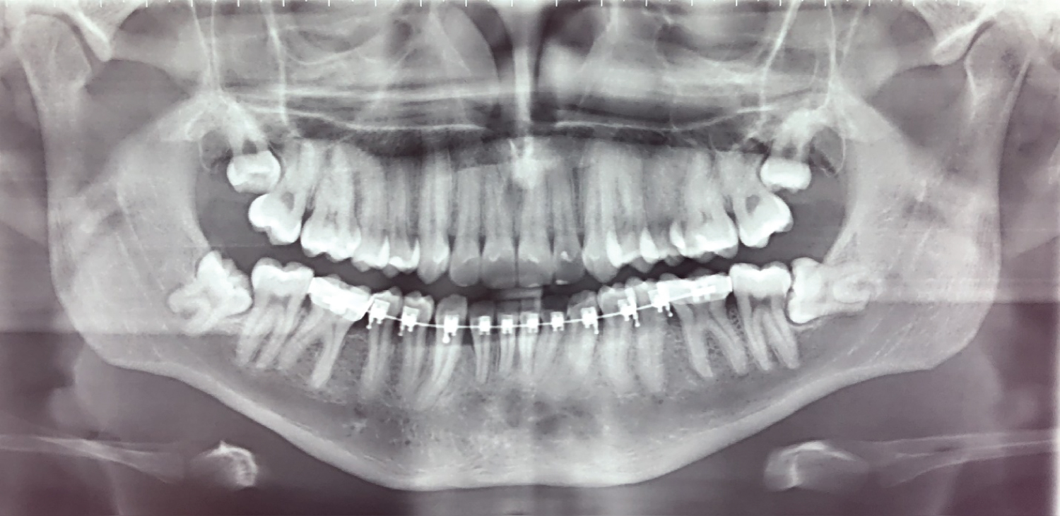
Third molar surgery is one of the most common surgical procedures performed by the oral and maxillofacial surgeon. Many articles regarding surgical technique and flap design follower third molar removal are available, although technical notes for impacted upper third molar surgery are uncommon. Classically, two incisions and a periosteum stripping are suggested for impacted upper third molar visualization and removal. We propose a less traumatic technique for impacted upper third molar surgery based on a unique vertical incision distally to second molar. By the fact that this technique only requires a single incision and minimal periosteum stripping, we truly believe that this technique can provide minor post-operative edema and faster patient recovery.
Keywords
Upper Third Molar, Surgery, Maxilla, Surgical approach
Introduction
Third molar surgery is one of the most common surgical procedures performed by the oral surgeon. Skill, experience and a systematic removal strategy are key steps for a clean and atraumatic surgery that provide better outcome and post-operative recovery [1]. Many articles regarding surgical technique and flap design for lower third molar removal are available, although technical notes for impacted upper third molar surgery are scares [2]. This fact can be attribute to less difficulty surgery, whilst severe complications have been reported on literature [3-5]. Classically, two incisions and a periosteum stripping is suggested for impacted upper third molar visualization and removal [6,7].
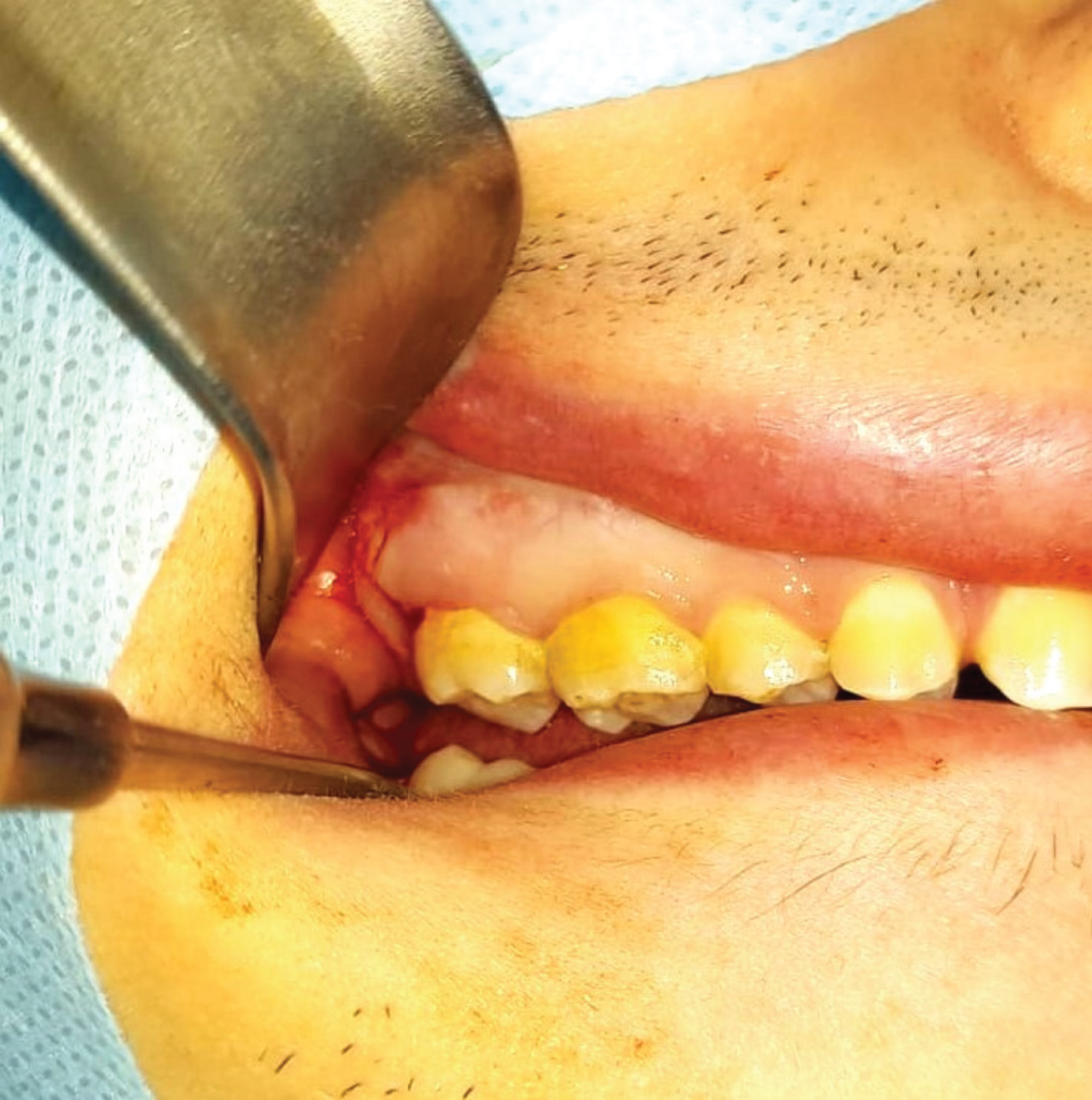
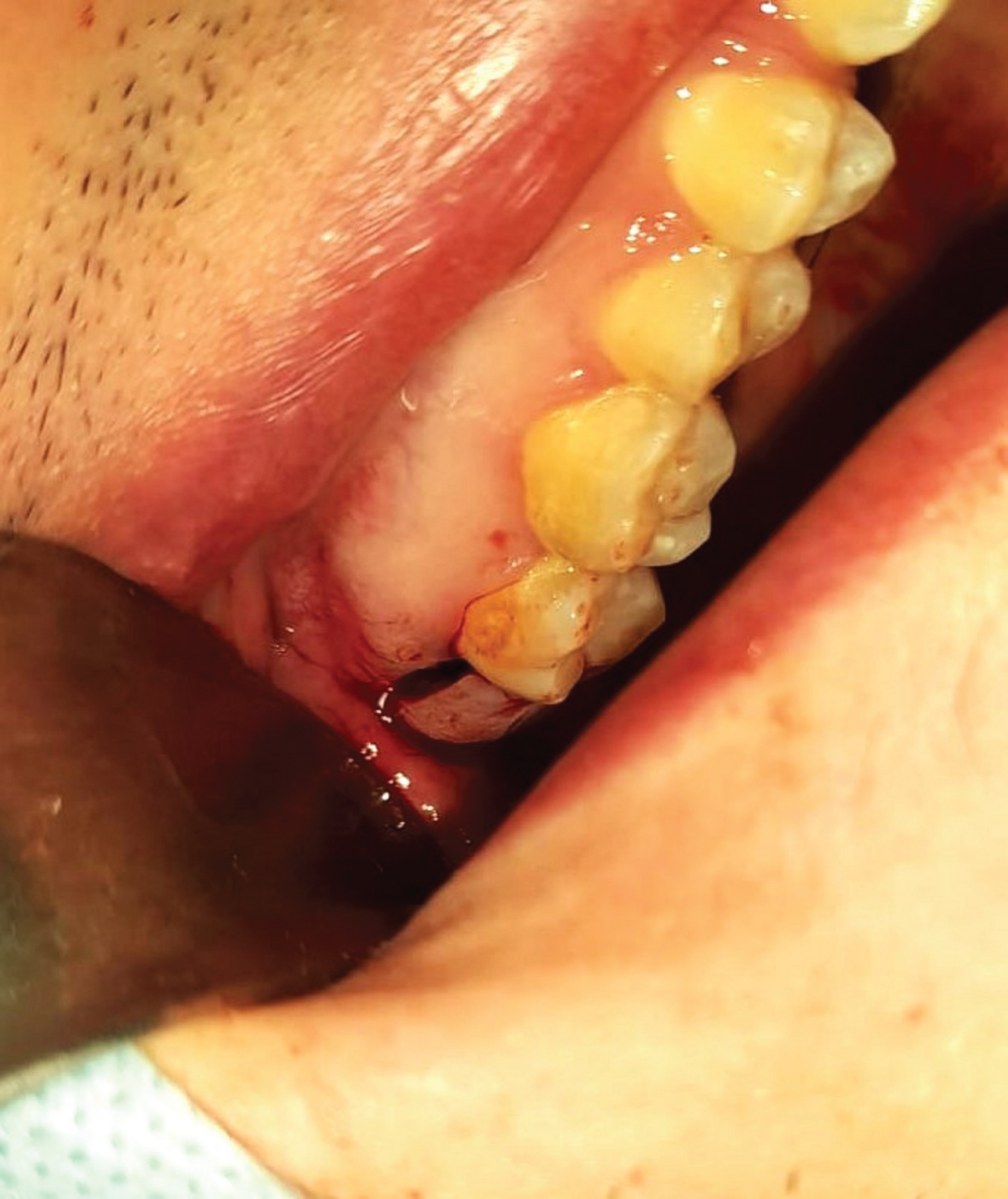
We propose a less traumatic technique for impacted upper third molar surgery based on a unique vertical incision distally to second molar, covering the attached gingiva up to the vestibule mucosa, reflecting the soft tissue flap enough to position the root elevator between the distal of the second molar and mesial of the upper third molar (Figure 1 and Figure 2). By the fact that this technique only requires a single incision and minimal periosteum stripping, and in the case of little visual access is still possible to make a second incision. Beyond that, in case of tuberosity fracture, bone maintenance might be possible due to periosteum vascular preservation, although more studies need to be done (Figure 3).
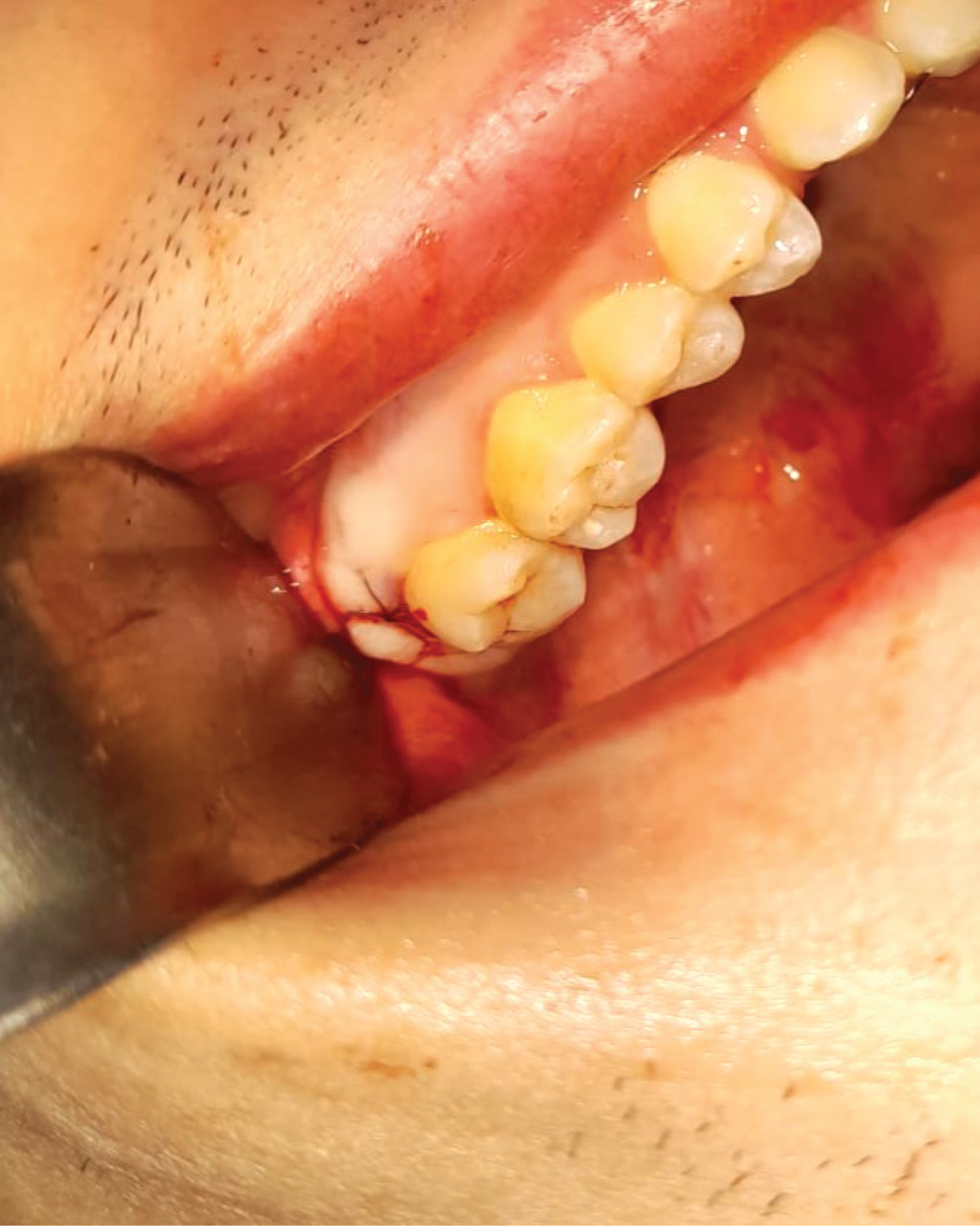
In conclusion, a single vertical incision can provide surgical access for most impacted upper third molar surgery. The suture is facilitated, because with one or two stitches the attached gingiva and free mucosa are closed (Figure 4). The fact that minimal periosteum stripping is required, a minor post-operative edema and faster patient recovery is expected.
Declarations
Funding
This research did not receive any specific grant from funding agencies in the public, commercial, or not-for-profit sectors.
Competing interests
No conflict of interest.
Ethical approval
No.
Patient consent
Patient consent has been obtained.
References
- Farish SE, Bouloux GF (2007) General technique of third molar removal. Oral Maxillofac Surg Clin North Am 19: 23-43.
- Carvalho RW, de Araujo Filho RC, do Egito Vasconcelos BC (2013) Surgical difficulty during removal of impacted maxillary third molars. J Oral Maxillofac Surg 71: 839-845.
- Selvi F, Cakarer S, Keskin C, et al. (2011) Delayed removal of a maxillary third molar accidentally displaced into the infratemporal fossa. J Craniofac Surg 22: 1391-1393.
- Gallagher J, Marley J (2003) Infratemporal and submasseteric infection following extraction of a non infected maxillary third molar. Br Dent J 194: 307-309.
- Lee JJ, Kok SH, Chang HH, et al. (2002) Repair of oroantral communications in the third molar region by random palatal flap. Int J Oral Maxillofac Surg 31: 677-680.
- Fragiskos D, Fragiskos (2007) Oral Surgery. Berlin: Springer, 156-159.
- Hupp JR, Tucker MR, Ellis E (2014) Contemporary Oral and Maxillofacial Surgery. (6 th edn), (Portuguese Edition) Elsevier, 379-440.
Corresponding Author
Elio Hitoshi Shinohara, DDS, PhD, Associate Surgeon, Special Oral Surgery Clinic, Cirurgia Bucal, Av Queiro Filho 1700, Torre B, cj 406, São Paulo, 05319-000, Brazil.
Copyright
© 2023 Shinohara EH, et al. This is an open-access article distributed under the terms of the Creative Commons Attribution License, which permits unrestricted use, distribution, and reproduction in any medium, provided the original author and source are credited.