Allogeneic Bone Graft for Stability of Inverted L Osteotomy Mandibular Ramus
Abstract
In corrections of dent facial deformities where the mandible is the problem, the inverted 'L' osteotomy may be a good option in specific cases, such as large mandibular advancements, condylar alterations, reoperations of the mandible, to increase the posterior facial height, among other situations. This technique was not much-used because the procedure used to be performed extra orally, leaving undesirable scars. We will describe the use of the technique via intraoral access, with allogeneic grafts and use of rigid internal fixation, exemplifying with some clinical cases.
Introduction
For corrections of dent facial deformities of mandibular origin, the sagittal osteotomy of the mandibular ramus is the most recommended technique. However, in some specific situations, the intraoral inverted 'L' osteotomy may be a good option. The extra oral inverted 'L' osteotomy is well described in the literature, but it can leave a cutaneous scar, which is undesirable for the patient. It is, therefore, a technique often forgotten by professionals. Some factors should be analyzed when choosing the technique to be used in the correction of mandibular deformities, like the versatility of the technique and absence of neurosensory changes. The technique should also allow the use of rigid internal fixation, offer surgical stability, and be easy to perform.
Bilateral sagittal osteotomy of the ramus (BSOR) is the most used technique due to its versatility and ease of fixation. However, there is a considerable risk of injury to the inferior alveolar nerve during the surgical procedure, in addition to the possibility of unfavorable fracture of the segments, change in the position of the mandibular condyles, especially torque, when using rigid fixation, and there is a discussion about the changes in the mandibular condyles during rotation movements of the maxillomandibular complex, suggesting that these movements could accelerate condylar resorption.
Such technique is recommended for some specific situations, e.g., reintervention after orthognatic surgery; large mandibular advancements (over 10 mm); mandibular asymmetries; corrections of the occlusal plane, especially in large counter clock wise rotations, which is considered a very unstable movement; patients with morphological changes in the condyles or with evident condylar resorption; treatment of ankylosissequelae; hemifacialmicrosomia; condyles with degenerative diseases; mandibular advancements in patients with masseteric hypertrophy, because such mandibles usually have almost no bone marrow, which makes the segment separation unpredictable; cases of open bite where the option of closing is through the mandible and where the recurrence rate remains low due to the use of bone grafts and rigid internal fixation. The intraoral inverted 'L' osteotomy (ILO) may be an alternative in these situations.
In this work, we will describe how we performed the intraoral inverted 'L' osteotomy in a series of clinical cases. We will demonstrate the versatility the technique, describing the sequence for its execution and comparing it with the findings in the literature.
Literature Review
Since its introduction as an option for correction of severe mandibular hypoplasia, the inverted 'L' osteotomy has not been the first choice for mandibular osteotomies, mainly due to the need for skin incisions, which may cause patient resistance. The technique was restricted to cases of greater severity where its application was essential, as in cases of ankylosissequelae, hemifacialmicrosomia, and large mandibular advancements.
However, with the development of the technique via intraoral access and the possibility of rigid fixation of the segments, the inverted 'L' osteotomy has been increasingly recommended. The inverted 'L' osteotomy has some benefits compared to the sagittal osteotomy of the ramus [1], such as: Improvement in ramus contour and mandibular angle, in patients with condylar changes; in planning that requires large counterclockwise rotations, making it difficult to perform the sagittal osteotomy, and greater projection of the lower facial third to improve aesthetics; when condylar stability is considered, especially in patients with degenerative joint disease or juvenile rheumatoid arthritis; to avoid torque in the mandibular condyles, preserving condylar function. The authors also point out that, with virtual planning, it is possible to position osteotomies in such a way as to facilitate the use of rigid fixation and accurately dimension the size and shape of the graft to be applied, greatly facilitating adaptation. In this study, not all cases had virtual planning, but we believe that its use facilitates the determination of the site and design of the osteotomy, the previous modeling of the graft and miniplates, and the choice of screws.
The inverted 'L' osteotomy [2] is indicated in the management of patients with the bird-face deformity of a severe class II malocclusion, high Frankfort-mandibular plane angle, and significant retrogenia, often associated with diminutive condyles and reduced posterior face height. The degree of mandibular advancement required and the potential for relapse is of greatest concern in these patients. The use of the sagittal technique is limited in these cases. We can also observe in these cases that there is greater stretching of the inferior alveolar nerve, contributing to paresthesia; there is poor contact of the bone stumps, even if extending the most anterior sagittal osteotomy; and there is no significant increase in the posterior facial height, because, in the sagittal osteotomy, a step that can make rigid fixation difficult is formed. When using an inverted 'L' osteotomy and placing grafts that collaborate for greater segment stability, there is a lower probability of relapse.
A study [3] investigated neurosensory disturbances in the lower lip and chin area in 21 patients who had undergone intra oral inverted 'L' osteotomy and 45 patients who had undergone sagittal osteotomy of the ramus. Through testing with electrical stimulation, the authors concluded that the inverted 'L' osteotomy offers less chance of injury to the inferior alveolar nerve when compared to the sagittal osteotomy. The authors also presented some advantages of the inverted 'L' osteotomy, namely: The posterior facial height is increased after mandibular repositioning; the condyle heads of the mandible are led automatically to a position where the masticatory muscles can retain their balance; and the period of intermaxillary fixation decreases considerably due to the possibility of fixing the segments. The highlight point observed in our sample was the passivity of the proximal segment in relation to the distal segment when maxillomandibular fixation was performed; the gap is formed very passively by muscular action. As for the postoperative fixation time, we recommended at least 2 weeks for fibrous tissue to collaborate in the stability of the osteotomies.
In a study [4] with 12 patients who underwent counterclockwise rotation of the mandible for correcting anterior open bite through modified inverted 'L' osteotomy, results showed good long-term stability. The authors concluded that, when the maxilla is well positioned, correcting anterior open bite with mandibular counterclockwise rotation is a good option. Inverted 'L' osteotomy and bone grafting can increase stability and decrease the chances of relapse.
Other authors [5] described a technique very similar to the one we used in our study. They point out that for the rigid fixation of the segments, the condylar segment is seated centrally in the fossa using gentle upward pressure from the area of the horizontal cut, the iliac crest bone graft is placed, and with a four-hole 'L' miniplate they perform the fixation of the segment. The authors used the inverted 'L' osteotomy in patients with hemifacialmicrosomia, condylar hypoplasia, temporomandibular joint ankylosis, and first and second branch arch syndrome.
There is no perfect technique; one must use good judgment in choosing the technique for each case, considering the anatomy of the mandibular ramus. In some authors' experience [6], mandibular rami associated with masseter hypertrophy show a large anteroposterior dimension and high density (almost no medullary bone), which increases the chances of undesirable fractures. In these cases, the inverted 'L' osteotomy is a good option. The authors suggest this technique be used when the mandible requires more than 12 mm of advancement. Also, in cases of reoperation, because the ramus probably suffered anatomical changes that may hamper the sagittal osteotomy. The inverted 'L' osteotomy is also indicated for correction of masseter hypertrophy, as shown by a study [7] that obtained satisfactory results in a sample of 15 patients.
As for the use of rigid fixation, there is no consensus on the best miniplate format, but bicortical screws are recommended. Virtual planning helps in the decision process, considering that each case will require different miniplates and screws due to the different anatomy of each patient and the extent and type of movement to be performed.
Among the difficulties observed in performing the technique via intraoral access are: Positioning and maintenance during the application of rigid fixation in the condylar (proximal) segment; and the difficulty in positioning and fixing the bone graft. The surgeon should have mastery of the use of the trocar, and the manipulation of the segments will depend on the learning curve.
The difficulty in fixing the segments [8] is a limitation for the popularization of the inverted 'L' technique, as there is a need for intermaxillary fixation for 4 to 6 weeks and difficulty in positioning the proximal segment. To control this problem, some authors suggest the use of a miniplate that anchors the proximal segment to the zygomatic buttress to stabilize the proximal segment and then complete the inverted 'L' osteotomy. With this maneuver, it is possible to keep the proximal segment in position and then perform the fixation of the inverted 'L' with miniplates and screws. In the cases performed in this study, we did not see the need to fix the proximal segment because, unintentionally, the segment remained in position, allowing fixation of the segments; however, this is a valuable resource to facilitate the application of rigid fixation.
Neurosensory changes caused by the sagittal osteotomy of the ramus make the inverted 'L' osteotomy a good option [9], especially in older patients who will undergo orthognathic surgery [10]. We agree with this fact, since the number of patients over 40 years of age who are candidates for orthognathic surgery has increased. Considering that the recovery of the inferior alveolar nerve is inversely proportional to the age of the patient, the inverted 'L' osteotomy is a good choice in this segment of patients.
Description of the Technique
The intra oral inverted 'L' osteotomy is applied both alone and in maxillomandibular surgeries, in normal or inverted sequence (i.e., starting from the mandible). In this sample of 10 cases, eight patients underwent bimaxillary surgery starting from the mandible and two patients underwent isolated mandibular surgery. The intraoral access is made very similar to that made for the sagittal osteotomy, but extending about 1 cm towards the mandibular ramus.
This facilitates the identification of the sigmoid notch and the placement of Bauer and Lavoisier retractors. After displacing the periosteum also in the lingual region to identify the lingula, and using a reciprocating saw, we start the horizontal osteotomy about 5 mm above the lingula, leaving about 5 to 7 mm below the posterior border of the mandibular ramus, and reaching vestibular cortical bone. With an oscillatory saw, we start the vertical ostetomy from the end of the horizontal osteotomy to the region of the mandibular angle. After separating the proximal and distal segments, we place the intermediate guide and perform the maxillomandibular fixation.
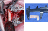
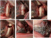
At the same moment, the gap between the segments is formed without the need to manipulate the distal segment. Depending on the extent of the mandibular movement, the gap will be larger or smaller, but in all patients we use allogeneic bone grafts (tibial cortical bone of the INTO (Instituto Nacional de Trauma e Ortopedia - Rio de Janeiro) bone bank). To fill the space, our choice is the region of the horizontal osteotomy, because this region offers the best view to adapt the graft. Once the graft is adapted, we model a miniplate, normally curved, with 6 to 8 holes, starting from the stump of the coronoid apophysis to the retromolar region. Using the trocar previously installed, we make the first perforation in the proximal segment (slightly pulling the segment, using a long Kely clamp, to facilitate the placement of at least two screws), hold the proximal segment by the miniplate, and fix the graft on the miniplate.
When possible, we put two screws on the graft. Next, we manipulate the proximal segment to passive position in the mandibular fossa. Then, we fix the screws to the distal segment (two screws). The same procedure is performed on the contra lateral side. Then, we release the maxillomandibular fixation to check the position of the mandible, joined together with the maxilla by the surgical guide, and suture with Vicryl 4-0. We recommend a period of intermaxillary fixation of 2 weeks. After this period, we recommend the continuous use of rubber bands for 30 days, removing them to eat (pasty food only) and perform oral hygiene, and gradually reduce the use of the bands in up to 60 days.
Cases Reports
Ten patients underwent intraoral inverted 'L' osteotomy, from 2010 to 2016. Four patients were female and six were male. Patients' ages at the time of surgery, clinical indications, and movements performed, follow-up, and relapses are shown in (Table 1). All patients are from the private clinic and signed the free and informed consent form, which includes authorization to use the images. Documents are signed by patients. Being granted exemption by the institutional review committee.
As we can see in this sample, the inverted 'L' technique has great versatility, since it was used in asymmetries, ankylosis, condylar alterations, reoperations, and to correct obstructive sleep apnea syndrome and open bite. The absence of torque in the mandibular condyles is a benefit of the technique. This reduces the possibility of condylar resorption, giving more passivity to the temporomandibular joint.
None of the patients in the sample had complaints in the region of the temporomandibular joint or reported any sensory changes in the inferior alveolar nerve. We had no difficulties in performing intra oral access in the inverted 'L' osteotomy. However, we had some difficulty (learning curve) in installing the trocar to perform the rigid fixation of the segments and of the graft. In the first three cases, we did not make any movements with the proximal segment, we just clamped the miniplate and the segment with a Kelly clamp to place the screws, which were difficult to visualize and insert. From then on, we started to model the graft with the proximal segment passive and the distal segment in position with the maxillomandibular fixation. After modeling the miniplate, we started to pull the proximal segment, facilitating the visualization of the coronoid process and the fixation of the screws to this segment and to the graft. Then, through the fixed miniplate, we positioned the proximal segment and fixed the screws to the distal segment more easily.
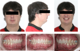
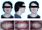
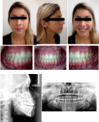
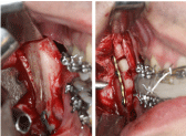
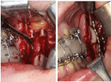
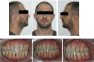
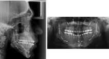
The grafts used in all cases, i.e., tibial cortical bone segments, were homologous grafts (bone bank). The choice of this graft was due to its rigidity, which favors stability with screws at the time of fixation, and to the amount needed to fill the gap and assist in stability, in addition to the lower morbidity compared with autologous grafts. The following cases exemplify the application of the technique. The first case was an OSAS (obstructive sleep apnea syndrome) patient using CPAP and undergoing orthodontic treatment (Figures 1a and 1b). In planning, we observed the need for maxillomandibular advancement and chin advancement. The result was a 21-mm pogonion advancement and a 17-mm mandibular advancement. We performed graft modeling for adaptation in the formed gaps (Figure 2), giving good stability to the mandible with rigid fixation (Figure 3a), as shown in the final radiographs and in the patient's facial and occlusal aspects (Figure 3b).
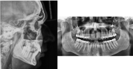
The second case was a patient who underwent surgery for bilateral ankylosis at 7 years of age and had hypo development of the mandible as a sequel (Figure 4a and Figure 4b). Figure 4b shows the initial radiographs. We waited for the patient to start orthodontic treatment and scheduled the inverted 'L' osteotomy. Specifically in this case, we removed the coronoid process on both sides. (Figure 5) shows the gap formed in the osteotomy when placing the mandible on the surgical guide. The objective was to give more mobility to the segment and improve mouth opening.
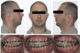
The positioning of the mandibular condyle is an important issue. In this sample of cases, we adopted movements of positioning the condyles with passivity, that is, using a long curved Kelly clamp, we slightly moved the proximal segment anteriorly and inferiorly to facilitate the placement of the screws in the coronoid process. Then, through the fixed miniplate, we passively manipulated this segment to the mandibular fossa and fixed the miniplate to the stump of the distal segment. No changes in condyle placement or symptoms were observed or reported after surgery, as seen in (Figure 6a) (face and occlusion), and in (Figure 6b) (postoperative radiographs).
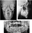
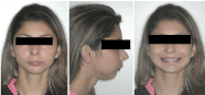
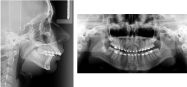
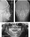
The third case was a patient who had already undergone orthognathic surgery of the maxilla and mandible (Figure 7a), but still had retrusion of the mandible and asymmetry on the left side, where we observed condylar alteration (Figure 7b). The patient, who is a wind instrument musician, said that he would not like to have his lip sensitivity altered again because this would impair his work. We performed the inverted 'L' osteotomy to avoid sensory changes and avoid tension in the condylar region, which could worsen the condition (Figure 8). So far, i.e., 5 years of follow-up, there has been no significant change in the result, both in facial and occlusal aspects (Figure 9a) and in radiographic follow-up (Figure 9b).
Final Considerations
The inverted 'L' osteotomy is a well-known technique, but the extra oral access limits its use. The aim of this article was to demonstrate the versatility of this type of osteotomy in different situations that occur in clinical practice. This technique is another tool that can be used in specific cases. The surgeon should have a certain amount of experience regarding the handling of the trocar, the positioning of the graft in the gap, and the use of rigid internal fixation. We believe that the advent of virtual planning, the manufacture of surgical guides that facilitate osteotomies, the modeling of grafts, and rigid fixation techniques can contribute to the popularization and evolution of the intraoral technique in the inverted 'L' osteotomy.
References
- Franco P B, Fareell B B (2016) Inverted L osteotomy: A new approach via intraoral access through the advances of virtual surgical planning and custom fixation. Oral and Maxillofacial Surgery Cases 2: 1-9.
- L Greaney, G Bhamrah, K Sneddon, et al. (2015) Reinventing the wheel: A modern perspective on the bilateral inverted 'L' osteotomy. Int J Oral Maxillofac Surg 44: 1325-1329.
- Kobayashi A, Yoshimasu H, Kobayashi J, et al. (2006) Neurosensory alteration in the lower lip and chin area after orthognathic surgery: Bilateral sagittal split osteotomy versus inverted l ramus osteotomy. J Oral Maxillofac Surg 64: 778-784.
- Aymach Z 1, Nei H, Kawamura H, et al. (2011) Evaluation of skeletal stability after surgical-orthodontic correction of skeletal open bite with mandibular counter clock wise rotation using modified inverted L osteotomy. J Oral Maxillofac Surg 69: 853-860.
- Song-Song Zhu, Ge Feng, Ji-Hua Li, et al. (2012) Correction of mandibular deficiency by inverted-L osteotomy of ramus and iliac crest bone grafting. Int J Oral Sci 4: 214-217.
- Medeiros P J, Ritto F (2009) Indications for the inverted-L osteotomy: Report of 3 cases. J Oral Maxillofac Surg 67: 435-444.
- Manganello-Souza L C, Oliveira A J, Maria Esther S, et al. (2000) Hipertrofia do Músculo Masseter Ver Soc Bras Cir Plas Sao Paulo 1: 45-54.
- Van Sickels J E, Tiner B D, Jeter T S (1990) Rigid fixation of the intraoral inverted'L'osteotomy. J Oral Maxillofac Surg 48: 894-898.
- McMillan B, Jones R, Ward-Booth P, et al. (1999) Technique for intraoral inverted 'L' osteotomy. Br J Oral Maxillofac Surg 37: 324-326.
- Hwang K, Nam Y S, Han S H (2009) Vulnerable structures during intraoral sagittal split ramus osteotomy. J Craniofac Surg 20: 229-232.
Corresponding Author
Joao Luiz Carlini, Associate Professor, Department of Surgery, Federal University of Parana, Curitiba, Brazil.
Copyright
© 2022 Carlini JL. This is an open-access article distributed under the terms of the Creative Commons Attribution License, which permits unrestricted use, distribution, and reproduction in any medium, provided the original author and source are credited.