Lipoma of the Parotid Gland: A Case Report and Review of Clinical Presentation, Method of Investigation and Management
Abstract
Lipomas are common mesenchymal tumours; however, their occurrence in the head and neck region is relatively rare and account for only 15% - 25% of all lipomas. Lipomas of the parotid region are even more uncommon, representing only about 1.0% -1.3% of all parotid tumours, and are often not included in the initial differential diagnosis of benign parotid tumours. Usually, a soft, non-tender, well-circumscribed tumour of the parotid gland in an elderly man is considered a Warthin's tumour unless proven otherwise by histopathological examination.
A rare case of a large lipoma of the parotid gland, which had been present for 15 years, is reported in a 63-year old male patient. The literature on clinical presentation, method of investigations and management of parotid lipoma is briefly reviewed.
Keywords
CT scan, FNAC, Lipoma, MRI, Parotid gland, Superficial parotidectomy, Ultrasound
Introduction
Lipomas are the most commonly encountered benign mesenchymal tumours. They are composed of mature fat cells and occur predominantly on the back, shoulder, and the abdomen [1,2]. Below the clavicle, lipomas are more common in obese female patients, usually those over 40 years of age; however in the head and neck region, the most commonly affected group are men in their fifth to seventh decade of life [3-5]. Indeed some studies have reported that men are more likely to be affected by head and neck lipoma by 52%, 62.5% and 68% respectively [6,4,7]. The occurrence of lipoma in the head and neck region is relatively rare, accounting for only about 15% to 25% of all lipomas [2,3,5,8,9] most of which occur subcutaneously in the posterior neck [4-6].
Parotid gland lipomas are very rare and are seldom considered in the initial preoperative differential diagnosis of benign parotid tumours [1,9-11]. Some authors report that lipomas of the parotid gland account for about 0.6% - 4.4% [3,12,13,14]. However, studies involving large series of patients report an incidence of 1.0% -1-3% of all parotid tumours [1,2,4,9,11,12]. Lipomatous lesions of the parotid occur in patients from 7 years to 72 years of age with a peak incidence between the 5th-6th decades of life and are 3-10times more common in males than females [2,3,8,9,13,15].
A rare case of a large left parotid gland lipoma of 15 years duration, which was initially diagnosed clinically as Warthin's tumor, is reported in a 63-year old male patient. The clinical presentation, diagnostic modalities and methods of treatment are briefly discussed.
Case Report
A 63-year old male Yemeni patient was referred to the Department of Oral & Maxillofacial Surgery by a General Surgeon, concerning a large, painless, gradually enlarging mass in the left parotid region of 15 years duration. There was no history of previous trauma or infection.
The patient's past medical history revealed him as a well-controlled diabetic and hypertensive, who was on glibenclamide, atenolol and nifedipine medication. He had undergone cholecystectomy 11 years earlier.
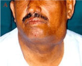
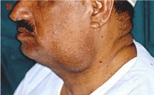
Physical examination showed a very large, soft, non-tender, well-circumscribed, mobile mass in the left parotid region posterior to the angle of mandible covered with normal over lying skin and measuring about 7cm x 4 cm (Figure 1A and 1B). The patient had grown a beard to conceal the mass. There was no alteration in facial nerve function, no palpable cervical lymph nodes, no trismus and salivary flow through the ipsilateral Stenson's duct was normal. The provisional clinical diagnosis considered, on account of his age, gender and the consistency of the swelling, was Warthin's tumor.
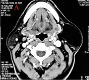
The CT scan revealed a well-circumscribed, smooth-walled, hypodense, non-enhancing lesion in the antero-superior aspect of the left parotid gland, measuring 5.1cm x 3.8 cm, with attenuation value of -120 Hounsfield Unit (HU) which corresponded to lipoma. There was mild compression of the external jugular vein but no cervical lymphadenopathy (Figure 2A).
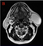
Magnetic resonance imaging (MRI) showed a well-demarcated mass involving the superficial lobe of the left parotid gland, which was hyperintense on T1-weighted and T2-weighted axial views as well as weak signals on fat-suppressed images (Figure 2B). Preoperative hematological investigation results were within normal range except for a fasting blood glucose level of 8.11mmol/l. Fine needle aspiration cytology (FNAC) of the parotid mass showed the presence of few adipose cells and no sign of malignancy.
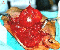
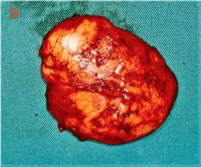
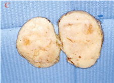
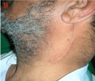
Under general anesthesia with orotracheal intubation, the flap was raised through the standard parotidectomy incision in the left preauricular region with cervical extension (classic Blair incision). When the parotid capsule was opened, a large lipomatous lesion was found entirely within the inferior aspect of the parotid gland. The soft, glistening, yellowish lump, measuring 7cm x 5cm x 3 cm, was excised by a combination of blunt and sharp dissection after preserving the buccal, marginal mandibular and cervical branches of the facial nerve (Figure 3A and 3B). Sectioned surgical specimen confirming the fat content of the mass is as shown in (Figure 3C). The wound was closed in layers under vacuum drainage. The post-operative wound healing was uneventful with no complications (Figure 4).
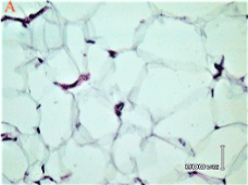
The histopathology examination was reported as "capsulated tumor with proliferation of lobules of adipose tissue separated by thin septa. There are focal congested blood vessels. No parotid tissue was seen in the capsule or in the tumor. The diagnosis is lipoma" (Figure 5). This confirmed the preoperative diagnosis provided by CT scan and MRI. The patient was discharged from clinic 2 years after surgery. It has been 15 years post-operatively and there is no sign of recurrence of the tumor as evident clinically and confirmed from a brain CT scan and MRI taken 13 years after surgery for an unrelated CNS ailment.
Discussion
Lipomas of the parotid can be classified, according to the site of occurrence, into paraparotid and intraparotid types. The paraparotid or superficial lipomas are those tumours that are found to be compressing the lateral surface of the parotid gland, adjacent to the parotid capsule or inside the capsule while the intraparotid or deep lipomas are tumours within the gland that are totally surrounded by salivary tissue [2,4,8,14,15,16]. However, there is a third group which involves both the superficial and deep lobes of the parotid gland [8,11,14, 15-17].
Clinical Presentation
Most parotid lipomas are found in relation to the superficial lobe [2,8,9,11,14] with very few reported deep lobe lipomas [1,4,5,8,10,12,18]. Indeed about 75% of parotid lipomas are located in the superficial lobe, 6.5% in the deep lobe and 16.5% are located in both lobes [19,20]. One study, however, reported more cases of deep lobe lipomas than the superficial ones by a ratio of approximately 2:1 [21]. A deep parotid lipoma may extend between the sternocleidomastoid and digastric muscles resulting in asymptomatic parotid swelling, or may extend to the parapharyngeal space with medial displacement of the lateral pharyngeal wall [15,17]. On both CT scan and MRI, the case in this report appeared to involve the superficial lobe of the left parotid gland.
Most lipomas of the parotid occur unilaterally even though bilateral ones have also been reported [2]. In unilateral cases, the majority of the lesions affect the left side more than the right [2,11,21] sometimes, by a ratio of 5:1 [2]. Janecka et al [8] in a review of 11 cases, reported more cases on the right than the left side while Walts & Perzik [3] in a review of 32 cases, reported an equal distribution in laterality. Lipomas of the parotid gland usually present as slow-growing, asymptomatic, non-tender, freely movable, soft masses [1,8,10,11] however, 30% of cases have been reported as indurated masses [19]. Occasionally, lipomas have been described as rubbery or hard in consistency [9,14,17,22]. Lipomas may present as focal encapsulated lesions or diffuse infiltrative masses. About 63% of parotid lipomas are of the focal variety [2,3]. Parotid lipomas have a benign presentation and are most often confused with Warthin's tumours or pleomorphic adenomas [3,4,9]. Clinically, a soft, non-tender and well-defined tumour in the parotid, in an elderly man, is normally considered a Warthin's tumour unless proven otherwise through histopathological examination [9]. Although they are generally asymptomatic, some parotid lipomas have been associated with facial paralysis [5,13,17], and pain [2,3,5]. Ethunandan et al [2]. (2006) reported that the painful parotid lipomas were all of the diffuse type and suggested that diffuse fatty infiltration of the glands may have led to distortion of the ductal architecture thus predisposing them to obstructive sialadenitis.
Methods of Investigation
Ultrasonography, high resolution CT scan or magnetic resonance imaging (MRI) are helpful in the further assessment and diagnosis of parotid lipomas. In the head and neck region, ultrasound shows lipomas as well-defined masses, hyperechoic to adjacent muscles however, others have also been described as isoechoic or hypoechoic so ultrasound is less specific than CT scan or MRI [16]. Compared to CT scan and MRI, ultrasonography is quick, easy, less costly and, with the use of high-frequency transducers, more suitable for imaging superficial lesions and therefore can be used as initial study. However, the soft tissue characterization seen in ultrasound is less specific than with CT scan or MRI and therefore where the mass is deep or difficult to identify on ultrasonogram, either CT scan or MRI is necessary as they provide the best tool for diagnosis and surgical planning [5]. They are the investigative tools of choice and are very accurate in preoperative diagnosis of lipoma. On CT scans, lipomas typically have characteristics of homogenous, well-encapsulated masses with few septations and specific negative attenuation with values ranging from -50 to -150 Hounsfield Units (HU) and with no contrast media enhancement [4,12,16,23]. MRI remains the best diagnostic imaging technique that can accurately diagnose lipoma preoperatively by comparison of signal intensity on T1-weighted and T2-weighted, as well as fat-suppressed images [4,5,12]. Lipomas usually show as strong signals on T1-weighted images and T2-weighted MR images and weak signals on fat-suppressed images [15,18,20]. The fat suppression sequence distinguishes these lipomatous tissues from other types of tumours. MRI, unlike CT scan, can also define the capsule of lipoma and its limit from normal adipose tissue by a black rim around the mass [14,16]. In addition, MRI is safe with few biological effects on the patient.
Although CT scan is three times cheaper, extremely valuable and as effective as MRI, if not more straightforward, the latter is preferable by many surgeons because of better imaging of soft tissue. The drawback for CT scan is that it exposes the patient to ionizing radiation, which could be avoided by using MRI [16,23]. The parotid lipoma in our report registered -120 HU on CT scan thus confirming lipoma. Magnetic resonance imaging (MRI) of the left parotid gland of the same patient showed a well-demarcated mass involving both the superficial lobe which was hyperintense on T1-weighted and T2-weighted views and showed weak signal on fat-suppressed images.
There is a general consensus among authors that one weakness in the current diagnostic imaging techniques in the diagnosis of tumours of fatty tissue is that, none of the imaging methods including CT scan and MRI can differentiate a lipoma from a liposarcoma. Such distinction can only be made with certainty by histopathological examination [4,16]. However, Kransdorf et al [24]. 2002, in a study of reliability of CT scan and MRI in distinguishing lipoma from liposarcoma, concluded that lipoma can be successfully distinguished from well-differentiated liposarcoma when it is completely composed of adipose tissue. Lipomas are benign tumours which are histologically similar to mature adipose tissue, but the presence of a fibrous capsule serves to distinguish them from simple aggregation of fat. The fibrous capsule shows as a black rim on MRI but is not observed on CT scan [16].
Fine needle aspiration cytology (FNAC) is very valuable in investigating parotid tumours (masses), but it is not helpful in lipoma. Although FNAC has been used with some success in parotid lipoma [1,7,8,10,23], other authors deem the technique unreliable for diagnosing a parotid lipoma due to a significant rate of false negative results in salivary gland tumours [4,5,18,23,25] even with experienced cytologist [25]. Moreover, fat cells from lipomas are identical to normal subcutaneous fat cells. Unlike normal adipose tissue, lipoma is not metabolized during starvation. When the diagnostic accuracy of MRI, CT scan and FNAC were compared in a small group of patients with parotid lipoma, it was concluded that CT scan and MRI are more reliable with 100% accuracy compared with 25% accuracy of FNAC in the diagnosis of lipoma [15]. Furthermore, FNAC may cause fibrosis or adhesion between the facial nerve branches and the lipoma capsule and this may increase the risk of facial nerve damage during surgery [5,15].
Management
The treatment of choice of parotid lipomas is surgical excision. The recommended surgical approach should be the same as for any other suspected benign parotid tumors with due consideration for the presence of facial nerve in the operation field. These include; superficial parotidectomy with nerve preservation [1,2,5,8,9,10,13,16,18], excision of the well-encapsulated lesion together with a rim of parotid gland tissue for periparotid or intraparotid lipomas [1,10,13,17], excision of the lipoma without superficial parotidectomy for those very close to the parotid capsule [11,26] and total conservative parotidectomy [2] for the deep parotid lipomas. Wu et al [5] and Ohyama et al. [22] recommend that when the deep lipoma is associated with the lower branches of the facial nerve, these branches should be meticulously dissected from the overlying superficial lobe instead of a formal superficial parotidectomy. Once the deep lipoma is removed, the raised superficial lobe should be repositioned. They believe that this technique helps to maintain better facial contour and avoid the need to excise redundant skin there by contributing to better post-operative aesthetic and functional results as well as preventing the occurrence of Frey's syndrome. Total parotidectomy is recommended especially for lipomas which involved both superficial and deep lobes, as an alternate treatment of choice [1,8,12,14]. Where lipoma occurs in a parotid gland whose parenchyma has been replaced by fat, it has been recommended that indigo carmine dye be injected through the Stenson's duct to stain the parotid gland to demarcate it clearly from the tumour thereby facilitating the excision of the latter [26].
The certainty with which CT scan and MRI, can establish the correct diagnosis of lipoma has led some authors to justify long-term clinical and imaging observation as a management option for small intraparotid lipomas [4,12] and clinically-static deep lipomas especially in patients reluctant to undergo surgery [1,12]. Surgical intervention in such patients may be reserved for those with cosmetic or pressure effects. Careful follow-up review for cases treated conservatively is necessary because malignant transformation has been reported [22]. If adequately resected, recurrence of a lipoma is less than 1% [9,11]. Re-excision of the recurrent lesion is curative [1,8]. Our patient has no recurrence 15 years after the surgery.
Conclusion
In the light of the case presented in this report, it is recommended that lipoma be included in the differential diagnosis of parotid tumours in elderly male patients. As the histopathology report confirmed, MRI and CT scan can be accurately used to diagnose lipoma on their own and therefore should be used, where it is available and affordable, to aid in the preoperative investigation of parotid tumours. While FNAC is useful in the diagnosis of some parotid tumours, it is rather unreliable when used in the diagnosis of parotid lipomas.
References
- Ozcan C, Unal M, Talas D, et al. (2002) Deep lobe parotid lipoma. J Oral Maxillofac Surg 60: 449-450.
- Ethunandan M, Vura G, Umar T, et al. (2006) Lipomatous lesions of the parotid gland. J Oral Maxillofac Surg 64: 1583-1586.
- Walts A E, Perzik S L (1976) Lipomatous lesions of the parotid area. Arch Otolaryngol 102: 230-232.
- Abd el-Monem M H, Gaafar A H, Magdy E A (2006) Lipomas of the head and neck: Presentation variability and diagnostic work-up. J Laryngol Otol 120: 47-55.
- Wu C W, Chi H P, Chiang F Y, et al. (2006) Giant lipoma arising from the deep lobe of the parotid gland. World J Surg Oncol 4: 28.
- Som P M, Scherl P M, Rao V M, et al. (1986) Rare presentations of ordinary lipomas of the head and neck: A review. AJNR Am J Neuroradiol 7: 657-664.
- Ahuja A T, King A D, Kew J, et al. (1998) Head and neck lipomas: Sonographic appearance. AJNR Am J Neuroradiol 19: 505-508.
- Janecka I P, Conley J, Perzin K H, et al. (1977) Lipomas presenting as parotid tumors. Laryngoscope 87: 1007-1010.
- Ryu J W, Lee M C, Myong N H, et al. (1996) Lipoma of the parotid gland. J Korean Med Sci 11: 522-525.
- Weiner G M, Pahor A L (1995) Deep lobe parotid lipoma; A case report. J Laryngol Otol 109: 772-773.
- Ansari M H (2006) Superficial lobe parotid gland lipoma. J Craniomaxillofac Surg 2006: 34: 47-49.
- Korentager R, Noyek A M, Chapnik J S, et al. (1988) Lipoma and liposarcoma of the parotid gland: High-resolution preoperative imaging diagnosis. Laryngoscope 98: 967-971.
- Kose R, Iynen I (2010) Lipoma of the superficial lobe of the parotid gland: A case report and review of the literature. Eur J Plast Surg 33: 215-218.
- Tilaveridis I, Kalaitsidou I, Pastelli N, et al. (2018) Lipoma of parotid gland: Report of two cases. J Maxillofac Oral Surg 17: 453-457.
- Paparo F, Massarelli M, Guiliani G (2016) A rare case of parotid gland lipoma arising from the deep lobe of the parotid gland. Ann Maxillofac Surg 6: 308-310.
- Tong K N, Seltzer S, Castle J T (2020) Lipoma of the parotid gland. Head Neck Pathol 14: 220-223.
- Srinivasan V, Ganesan S, Premachandra D J (1996) Lipoma of the parotid gland presenting with facial palsy. J Laryngol Otol 110: 93-95.
- Ulku C H, Uyar Y, Unaldi D (2005) Management of lipomas arising from deep lobe of the parotid gland. Auris Naris Larynx 32: 49-53.
- Fakhry N, Michel J, Varoquaux A, et al. (2012) Is surgical excision of lipomas arising from the parotid gland systematically required? Eur Arch Otolaryngol 269: 1839-1844.
- Rehal S S, Alibhai M, Perera E (2017) Lipoma of the parotid after trauma: A case report and review. Br J Oral Maxillofac Surg 55: e5-e6.
- Furlong M A, Fan burg-Smith J C, Childers E L B (2004) Lipoma of the oral and maxillofacial region: Site and sub classification of 125 cases. Oral Surg Oral Med Oral Pathol Oral Radiol Endod 98: 441-450.
- Ohyama Y, Shigematsu H, Kwawamoto Y, et al. (2012) A case of deep lipoma. Journal of Oral and Maxillofacial Surgery Medicine, and Pathology 24: 132-135.
- Dellaportas D, Mantzos D S, Theodosopoulos T, et al. (2016) Parotid gland lipoma: An unusual entity. Chirugia 111: 64-66.
- Kransdorf M J, Bancroft L W, Peterson J J, et al. (2002) Imaging of fatty tumors: Distribution of lipoma and well-differentiated liposarcoma. Radiology 224: 99-104.
- Layfield L J, Glasgow B J, Goldstein N, et al. (1991) Lipomatous lesions of the parotid gland: Potential pitfalls in fine needle aspiration biopsy diagnosis. Acta Cytol 35: 553-556.
- Sato T, Terada C, Ide S, et al. (2021) A case of lipoma of the parotid gland: Distinguishing the tumor from the fatty parotid gland by the injection of indigo carmine into Stenson's duct for appropriate enucleation. Journal of Oral and Maxillofacial Surgery, Medicine, and Pathology 33: 416-419.
Corresponding Author
Braimah Ramat Oyebunmi (BDS, FAOCMF, FMCDS, FWACS), Department of Oral and Maxillofacial Surgery, Specialty Regional Dental Center, Najran, Kingdom of Saudi Arabia.
Copyright
© 2021 Daniels JS. This is an open-access article distributed under the terms of the Creative Commons Attribution License, which permits unrestricted use, distribution, and reproduction in any medium, provided the original author and source are credited.

