Prevalence of Keratoconus in Patients Attending Outpatient Clinic in Shahid Aso Eye Hospital for Refractive Problem
Abstract
Background : Keratoconus is characterized by the thinning of the cornea and irregularities of the cornea’s surface. Keratoconus is a common ophthalmic disease worldwide especially in adulthood. Keratoconus generally begins at puberty and progresses into the mid-30s. This condition is more common in the Middle East than the rest of the world. In Iraq and Kurdistan the rate of this disease has increased.
Objective : To determine the prevalence of keratoconus in patients attending Shahid Aso Eye Hospital with refractive error between January 2024 to June of the same year.
Method : Our prospective cross sectional research study examined the clinical data in recording files( in which Corneal Tomography measurements were used to help detecting keratoconus) were reviewed, (305)patients among them (169) were females and (136) were males consulting Shaheed Aso eye hospital for correcting their refractive errors during the period between Jan till June, 2024.
Result : Among (305) patients with refractive errors in all age groups, (92) patients (30.2%) have been diagnosed as keratoconus.
Conclusion : The prevalence of keratoconus was higher among younger patients. Female were the dominant gender.
Keywords
Keratoconus, Corneal thickness, Pentacam, Allergy, Refractive Error
Introduction
Definition
Keratoconus is a progressive non-inflammatory corneal ectatic disorder characterized by thinning and protrusion of the cornea, resulting in irregular astigmatism and visual impairment [1].
The term keratoconus is derived from the Greek words kerato (cornea) and konos (cone), reflecting the conical shape that the cornea assumes in this condition [2]. The exact etiology of keratoconus remains unknown, but it is believed to involve a complex interplay of genetic, environmental, and biomechanical factors [3].
Keratoconus is a relatively rare condition and typically present in adolescence or early adulthood with a peak onset between the ages of 10 and 25 years [4]. It affects both genders and is more common in certain ethnic groups, such as south Asian and middle eastern populations [5].
Histopathology
Histopathological findings in keratoconus include break in bowman's layer, stromal thinning, collagen fibril disorganization, and corneal ectasia. The risk factors for the condition include family history of keratoconus, eye rubbing [2] and allergy.
Mechanical trauma from rubbing leads to trauma to the keratocytes and increases pressure in the eyes [3].
Wearing contact lenses also causes corneal micro trauma that is linked to keratoconus [6]. There is evidence of environmental and genetic involvement [7,8], while the hereditary pattern of keratoconus is unpredictable. Several loci associated with autosomal dominant inherited keratoconus has been identified [9].
Classification
Several classification systems have been proposed for keratoconus, based on various criteria such as severity, progression, and topographic patterns [10].
The most widely used classification system is the Amsler-Krumeich classification, which categorizes keratoconus into four stages based on the degree of corneal thinning, protrusion, and scarring [11,12]. Other classification systems, such as the Belin-Ambrosio Enhanced Ectasia Display, have been developed to provide a more comprehensive assessment of corneal ectasia [13].
Diagnosis
For diagnostic evaluation, corneal topography is a valuable tool for detecting subclinical keratoconus and monitoring disease progression. Many quantitative keratograph indices have been developed for screening keratoconic patients including corneal steepening, astigmatism and irregular astigmatism, pachymetry and other parameters aid in detecting keratoconus suspect to the diagnosis and management of keratoconus [14].
Clinical presentation
The clinical presentation of keratoconus can vary widely,but common signs and symptoms include progressive myopia, irregular astigmatism [15] glare, halos and decreased visual acuity, particularly in low light conditions [16].
Early signs such as scissors reflex during retinoscopy, fleischer ring near the base of the cone, fine striation (Vogt lines), and munson’s sign,where corneal protrusion causes angulation of lower lid on down gaze. The diagnosis of keratoconus is typically made based on a combination of clinical findings, corneal topography and corneal tomography [17].
Management and treatment
The management of keratoconus typically involves a combination of spectacles, contact lenses, and surgical interventions.
Spectacles and rigid gas permeable contact lenses are often used to correct refractive errors and improve visual acuity in early stages of keratoconus. In more advanced cases, surgical interventions
Such as corneal collagen cross-linking, intrastromal corneal ring segments, and keratoplasty may be necessary to stabilize or improve visual function [18-20].
Recent advances in contact lens design, such as scleral and hybrid lenses, have also provided new options for managing keratoconus [21].
Research Methodology
This is prospective cross sectional research study examined the clinical data in recording files( in which Corneal Tomography measurements were used to help detecting keratoconus) were reviewed, (305)patients among them (169) were females and (136) were males consulting Shaheed Aso eye hospital for correcting their refractive errors during the period between Jan till June, 2024.
For each patient full medical and ocular history were taken including:family history, history of allergy , history of rubbing and presence of other medical problems. Through ocular examination was taken including visual acuity and slit lamp examination. Then pentacam examination was performed and results documented.
Ethical consideration regulated by the ministry of higher education and ministry of health were observed during the conduction of the study. Informed consent was taken from the patients clarifying that this data are going to be used by the research team and willingness of patient to share his or her documented information for the research purpose were clear in the consent.
Data collected and analysed using software SPSS statistical package for the social science analysis. Chi-square test and ANOVA to identify correlation between variables at the statistical level of 0.05.
Results
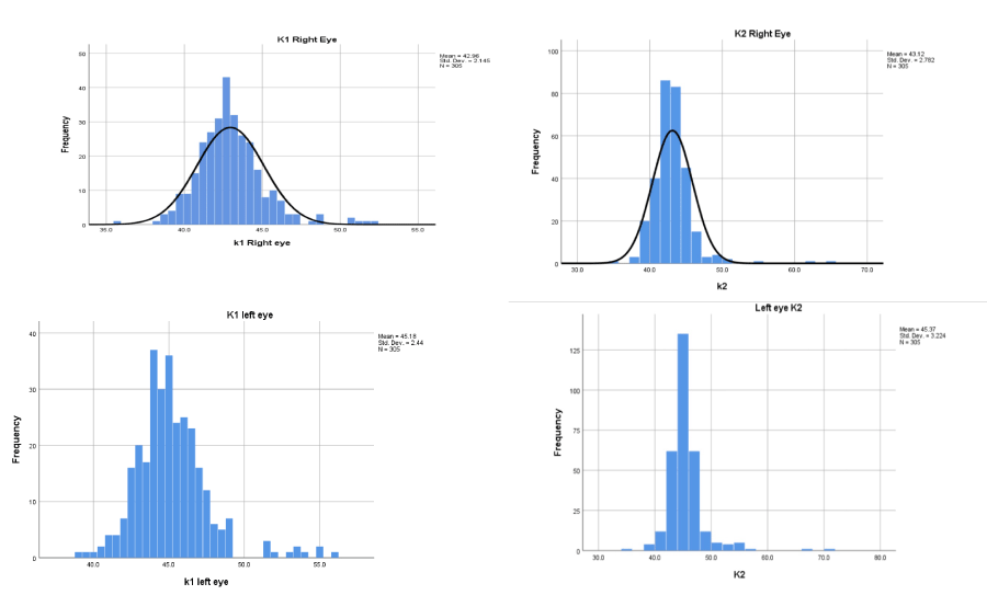
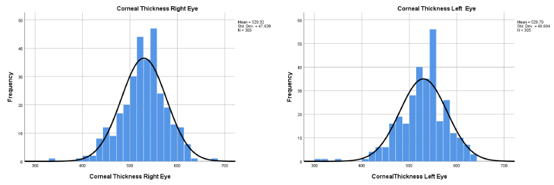
A total of 305 patients 610 eyes were included in this study. Of these, 92 patients (30,2%) had keratoconus and 213 patients had no Keratoconus (69.8). These patients had a mean age of 23.41 (range: 9-41 years) and 169 (55.4%) were females 136 (44.6%) were males (Figure 1 and 2) and (Table 1).
Discussion
The aim of this study is to determine prevalence of keratoconus patients attending Shaheed Aso hospital from January till June 2024.the study includes 305 patients, among them 30.2% have keratoconus while in Saudi Arabia among 548 patients 24% have keratoconus .
In our study eye itching, allergy and positive family history for keratoconus were the most risk factors for causing keratoconus, but our findings of positive family history was 31.1% in comparison with those in Middle East in Saudi Arabia 16% and Lebanon 12.1% [22,23].
We detect keratoconus through history taking, refraction test, slit lamp examination, and then Tomography for confirming the diagnosis. Early diagnosis of keratoconus increase the quality of vision and life through early management and prevent progression of disease by using CXL (cross linking).
The prevalence of keratoconus in our study is 30.2% while it was 0.02% in Saudi Arabia. Females were dominant 55.4% which was matched with the study done in Iraq 61.1% [24].
Conclusion
Current research study found that the prevalence of keratoconus was higher among patients between 12-40 years old, 30.2% (92 patients per the total 305 patients) the mean age was 23.41, keratoconus is higher in younger than the older patents so There is a significant correlation between development of keratoconus and age of participants readings as the p value is 0.00 (using one way ANOVA). with females being the dominant gender, the females were 55.4% and the males were 44.6%,so There is no significant correlation between keratoconus diagnosis and sex, p value is 0.9 using chi- square test. There are 50 patients (16.4%) having history of allergy so There is a significant correlation between diagnosis of keratoconus and history of allergy as the p value is 0.00 using Chi -square test, and 94 (30.8%) patients have history of itching so there is a significant correlation between keratoconus diagnosis and itching history p value is 0.00 using Chi-square test, 95 patients (31.1%) hae positive family history there is a significant correlation between diagnosis of keratoconus and family history as the p value is 0.00 using Chi -square test the mean K142.95, K2 43.11 of right eye and mean K1 45.17, mean K2 45.36 of left eye there a significant correlation between diagnosis of keratoconus and K1, K2, reading of right and left eye as ,the p value in all the instances is 0.00(using one way ANOVA). Regarding mean corneal thickness of right eye is 529.32, standard deviation 47.63 and the mean corneal thickness of left eye is 529.79 with standard deviation 49.69 so there is a significant correlation between diagnosis of keratoconus and corneal thickness of right and left eye as the P value is 0.00 (using one way ANOVA).
Keratoconus is a progressive corneal disorder that can lead to visual impairment and irregular astigmatism. Early detection through corneal tomography measurements is crucial for timely management and treatment. Further research and awareness are needed to better understand the etiology and risk factors associated with keratoconus. Effective management strategies, including spectacles, contact lenses, and surgical interventions, can help stabilize or improve visual function in patients with keratoconus.
References
- Valdez-García JE, Sepúlveda R, Salazar-Martínez JJ, et al. (2014) Prevalence of keratoconus in an adolescent population. Revista Mexicana de Oftalmología 88: 95-98.
- Netto EA, Al-Otaibi WM, Hafezi NL, et al. (2018) Prevalence of keratoconus in paediatric patients in Riyadh, Saudi Arabia. Br J Ophthalmol 102: 1436-1441.
- Papali’i-Curtin AT, Cox R, Ma T, et al. (2019) Keratoconus prevalence among high school students in New Zealand. Cornea 1382-1389.
- Grzybowski A, Mcghee CNJ (2013) The early history of keratoconus prior to Nottingham’s landmark 1854 treatise on conical cornea: A review. Clin Exp Optom 96: 140-145.
- Ertan A, Muftuoglu O (2008) Keratconus clinical finding according to different age and gender groups. Cornea 27: 1109-1113.
- Armstrong BK, Smith SD, Romac Coc I, et al. (2021) Screening for keratoconus in a high-risk adolescent population. Ophthalmic Epidemiol 28: 191-197.
- Maguire LJ, Singer DE, Klyce SD (1987) Graphic presentation of computer-analyzed keratoscope photographs. Arch Ophthalmol 105: 223-230.
- Wilson SE, Klyce SD, Husseini ZM (1993) Standardized color-coded maps for corneal topography. Ophthalmology 100: 1723-1727.
- Barbara A (2012) Textbook on Keratoconus: New Insights. Jaypee Brothers 224.
- Rathi VM, Mandathara PS, Dumpati S (2013) Contact lens in keratoconus. Indian J Ophthalmol 61: 410-415.
- Grisevic S, Gilevska F, Biscevic A, et al. (2020) Keratoconus progression classification one year after performed crosslinking method based on ABCD keratoconus grading system. Acta Inform Med 28: 18-23.
- Ucar M, Cakmak HB, Sen B (2016) A statistical approach to classification of keratoconus. Int J Ophthalmol 9: 1355-1357.
- Bamdad S, Sedaghat MR, Yasemi M, et al. (2020) Sensitivity and specificity of belin ambrosio enhanced ectasia display in early diagnosis of keratoconus. J Ophthalmol 2020.
- Martinez-Abad A, Piñer DP (2017) New perspectives on the detection and progression of keratoconus. J Cataract Refract Surg 43: 1213-1227.
- Estrada AV, Díez PS, Alió JL (2016) Keratoconus grading and its therapeutic implications. Keratoconus 177-184.
- Abe RY, Medeiros FA, Davi MA, et al. (2019) Psychometric properties of the Portuguese version of the National Eye Institute visual function questionnaire-25. PLoS ONE 14: e0226086.
- Toprak I, Vega A, Del Barrio JLA, et al. (2021) Diagnostic value of corneal epithelial and stromal thickness distribution profiles in forme fruste keratoconus and subclinical keratoconus. Cornea 40: 61-72.
- Tasçi YY, Taslipinar G, Eyidogan D, et al. (2020) Five-year long-term results of standard collagen cross-linking therapy in patients with keratoconus. Turk J Ophthalmol 50: 200-205.
- Nosé W, Neves RA, Burris TE, et al. (1996) Belfort Intrastromal corneal ring: 12-month sighted myopic eye. J Refract Surg 12: 20-28.
- Gadhvi KA, Romano V, Cueto LFV, et al. (2019) Deep anterior lamellar keratoplasty for keratoconus: Multisurgeon results. Am J Ophthalmol 201: 54-62.
- Rubinstein MP, Sud S (1999) The use of hybrid lenses in management of the irregular cornea. Cont Lens Anterior Eye 22: 87-90.
- Assiri AA, Yousuf BI, Quantock AJ, et al. (2005) Incidence and severity of Keratoconus in asir province, Saudia Arabia. Br J Ophthalmol 189: 1403-1406.
- Waked N, Fayad AM, Fadalallah A, et al. (2012) Keratoconus screening in Lebanese students population J Fr Ophtalmol 35: 23-29.
- Qasim KF, Shahad A (2017) Prevalence of refractive errors in patients with keratoconus among sample of iraqi population. J Ophthalmol 2: 1-8.
Corresponding Author
Kosar Ali Rashid, Consultant ophthalmologist in shahid Aso eye hospital, Sulaimanyah, Iraq.
Copyright
© 2025 Rashid KA, et al. This is an open-access article distributed under the terms of the Creative Commons Attribution License, which permits unrestricted use, distribution, and reproduction in any medium, provided the original author and source are credited.


