Ruptured Middle Cerebral Artery Aneurysms: Retrospective Study and Multivariate Analysis of 105 Patients Treated by Surgical Clipping
Abstract
Objective of the study
We analyze in this study only patients with surgically treated ruptured aneurysms in order to identify statistical significance of each predictive factor in terms of outcome of patients with ruptured MCAAs.
Materials and methods
In this retrospective study, we analyzed 105 cases of ruptured MCAAs, admitted from January 2001 to December 2015 at Neurosurgical Department of Umberto I University Hospital of Rome, Italy. Predictive factors evaluated are: Patient's features (age, sex, co-morbidities), aneurysmal location (proximal, bifurcation or distal) and size of aneurysmal dome (small, large or giant); surgical timing (ultra-early, early, delayed), and Intracerebral Hemorrhage (ICH) volume. For each parameter we calculated mean and standard deviation, covariance and relation coefficient (through the linear regression model).
Results
The clinical evaluation of patients assessed through the World Federation of Neurological Surgeons (WFSN) grading scale, that is 5 for 37 patients (35.3%), 4 for 28 patients. In 40% of cases the maximum sac diameter was between 7 mm and 12 mm, while in 67% of the cases the aneurysms concerned the bifurcation of the middle cerebral artery. ICH was associated in 57 cases (54.3%). As far as outcome is concerned, at 3 months, 32 patients (30.47%) had a favourable outcome, while 73 (69.52%) patients had not favourable outcome. To one year, 46 patients (43.8%) had favourable outcomes, while 59 patients (56.19%) had not favourable outcome. The mean outcomes as mean mRS are significantly less favourable in patients with ICH.
Conclusion
In MCAAs patients, the presence of ICH strongly affects the outcome with a marked increase in mortality and morbidity. Surgical timing significantly influences the outcomes and ultra-early surgery should always be taken into account.
Keywords
Middle cerebral artery, Aneurysms, ICH, Outcome, Middle cerebral artery aneurysms, Ultra-early surgery, Early surgery, Delayed surgery
Abbreviations
MCAAs: Middle Cerebral Artery Aneurysms; SAH: Subarachnoid Haemorrhage; ICH: Intracerebral Haemorrhage; WFSN: World Federation of Neurological Surgeons; mRS: Modified Rankin Scale; IVH: Intraventricular Haemorrhage; GCS: Glasgow Coma Scale; OLS: Linear Regression Model; MCAbifAs: Bifurcation Aneurysms; CSF: Cerebrospinal Fluid
Introduction
Middle Cerebral Artery Aneurysms (MCAAs) represent 20% of all intracranial aneurysms. Most of them (80%) are located on the sylvian bifurcation, the seat of hemodynamic turbulence flow [1,2]. The rupture of this aneurysm causes Subarachnoid Hemorrhage (SAH), with frequently associated Intracerebral Hemorrhage (ICH). The associated ICH seems to be a significant prognostic factor, and usually presents as a temporal ICH, or less frequently as intra-sylvian hematoma. Best management strategy of MCAAs is still discussed: The rapid evolution in endovascular coiling or stenting techniques achieved similar results to those of the surgical clipping: Choosing the best treatment indication randomly changes. We analyze in this study only patients with surgically treated ruptured aneurysms in order to identify statistical significance of each predictive factor in terms of outcome of patients with ruptured MCAAs.
Materials and Methods
In this retrospective study, we analyzed 105 cases of ruptured MCAAs, admitted from January 2001 to December 2015 at Neurosurgical Department of Umberto I University Hospital of Rome, Italy. Within these 105 patients, there were 48 patients presenting with aneurysmal rupture and Subarachnoid Hemorrhage (SAH) and 57 who presented with associated ICH. Inclusion criteria are: 1) All adult patients surgically treated for ruptured MCAAs; 2) With or without aneurysm localization and 3) With or without re-bleeding. Exclusion criteria are: 1) Aneurysms treated by endovascular embolization/stenting; 2) Patients admitted for another aneurysm location than MCA; 3) Post-traumatic aneurysmal rupture 4) Unruptured aneurysms. The diagnostic techniques used have been CT angiography, and/or preoperative angiography. Predictive factors evaluated are: Patient's features (age, sex, co-morbidities), aneurysmal location (proximal, bifurcation or distal) and size of aneurysmal dome (small, large or giant); surgical timing (ultra-early, early, delayed), and ICH volume.
Clinical evaluation scale
The clinical grade have been assigned to each patient at admission time according to the World Federation of Neurological Surgeons (WFNS) grading score, basing on Glasgow Coma Scale (GCS) and presence or absence of focal deficits. Each patient have been classified in terms of outcome as having either a good recovery including moderate disability (a favorable outcome) or a poor recovery including severe disability, or vegetative state (not favorable outcome). Functional outcome of patients after 3 months and after 1 year was evaluated using the Modified Rankin Scale (mRS). It was judged as favorable (mRS = 1-3) and not favorable (mRS = 4-6).
Severity evaluation scale
In this study we have used Fisher scale to evaluate severity of SAH and ICH score which allows risk stratification on presentation with ICH [3-5]. These models usually include criteria related to neurological condition (GCS) and neuroimaging findings (ICH volume, IVH, location).
Aneurysmal features
The size of the aneurysmal dome was measured using the largest diameter measurement based on the long or perpendicular axis of the dome. The aneurysm sizes have been divided into four categories: 1) Smaller than 7 mm; 2) Between 7-12 mm; 3) 12-24 mm; 4) > 24 mm. MCAAs have been classified into 3 groups according to the location of the aneurysm neck: Proximal (n = 16), bifurcation (n = 70), and distal aneurysms (n = 19).
Criteria for assignment to surgical treatment
Historically, middle cerebral artery aneurysms have been considered surgical lesions. This type of aneurysms is not frequently accounted ideal for endovascular coiling for their unfavorable anatomic features (wide neck and partial incorporation of one or both M2 branches). However, endovascular techniques for aneurismal treatment have significantly expanded [6].
In our series, the choice of surgical treatment was based on physician on-call schedule evaluation in emergencies, indeed 54.28% of patients have been treated in ultra-early surgery, 33.33% in early surgery, and only 12.28% in delayed surgery. Criteria for assignment to surgery have been the presence of ICH and wide neck aneurysm.
Surgical procedure
Through a pterional craniotomy, wider than the ordinary approach used for aneurysms, obliteration of MCAA was performed via a trans-sylvian approach using standard microsurgical dissection [7]. In 24 patients ICH evacuation was accomplished after aneurysmal clipping, on the other hand in 33 patients it was performed before aneurysmal clipping; the only aneurysmal clipping was performed in 44 patients without ICH. In case of brain swelling, a duroplasty was performed and the bone flap was not replaced.
Statistical analysis
We studied all collected data with the programs Excel and MATLAB. For each parameter we calculated mean and standard deviation and when necessary Spearman's rank correlation coefficient, covariance and relation coefficient (through the linear regression model).
Results
The mean age of the patients included in our study was 59 ± 13 years, with a very large age range, from 22 to 96 years. Out of 105 patients, 60 are female (62%) and 45 male (38%). Table 1 show the WFNS grade, Fisher scale and ICH score at the entrance of subjects included in the study. An associated ICH is present in 57 patients (54.3%). Most of patients (23) present an ICH score of 2, 12 patients have an ICH score = 3, 11 patients have ICH score = 4, 6 patients have ICH score of 5 and 5 patients present ICH score of 1. In 57 patients (54.3%) the CT Angiography shows Fisher scale grade IV. The clinical evaluation of patients assessed through the WFSN grading scale; is 5 for 37 patients (35.3%), 4 for 28 patients. Regarding the aneurysmal diameter and the localization, in the 40% of cases the maximum diameter was between 7 mm and 12 mm, while in 67% of the cases the aneurysm concerned the bifurcation of the middle cerebral artery. About surgical timing, in 57 patients (54.28%) we performed evacuation of hematoma and clipping of the aneurysm in ultra-early surgery (< 12 h) from the onset of symptoms, in 35 patients (33.33%) in early surgery (< 48 h) and in 13 patients (12.38%) in delayed surgery (> 48 h). The 8.7% of aneurysms re-bleached after the first event in ultra-early surgery patients, whereas in early-surgery and in delayed surgery patients the re-bleeding occurred in 17.14% and 15.30% respectively. Concerning outcome, at 3 months, 32 patients (30.47%) had a favourable outcome, meaning mRS ≤ 2, while 73 patients (69.52%) had not favourable outcome, meaning mRS ≥ 3. In one year, 46 patients (43.8%) had favourable outcomes, while 59 patients (56.19%) had not favourable outcome. The mortality rate at 3 months and 1 year after bleeding was 14.28% (15 patients) and 20% (21 patients) respectively, reported in Table 2. In addition, the mean outcomes as mean mRS are significantly less favourable in patients with ICH (see below). In Table 3 are summarized all data analyzed with their statistical significance. It is noted that among the parameters we analyzed, those that were statistically significant (p-value < 0.05) are the presence or absence of ICH, age of the patients, the clinical status at the entrance (WFNS grade), the surgical timing and the severity of SAH (Fisher scale). The diameter and seat of the aneurysm does not appear to influence outcome. In our study, the analysis of the latter two parameters in relation to surgical outcome was not statistically significant.
ICH and outcome
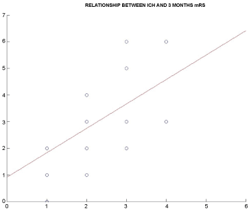
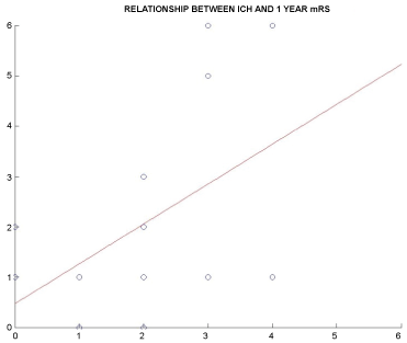
The mean 3 months and 1 year mRS in patients with ICH was respectively 4.3 ± 0.72 and 3.6 ± 0.81. The mean 3 months and 1 year mRS in patients without ICH was 3.3 ± 0.64 and 2.6 ± 0.73 respectively. In patients with ICH, the mean ICH score (see Table 1) was 2.82 ± 1.13. We also estimated the correlation (ρ) (values between -1 and 1) between the ICH score and three-months and one-year results, obtaining the following values: 0.73 at 3 months and 0.60 at one year, both positive. The covariance σ (ICH score, mRS) at three months and one year was 1.44 and 1.13 respectively, both positive to indicate that the two values positively combined. In last through a Linear Regression Model (OLS) we estimated the variable regression coefficient ICH score against mRS (and therefore outcome) obtaining the following results: 0.93 to three months and 0.81 to a year. This indicates that an increase of 1 ICH-score units involves an expected increase in the score in the MRS scale of 0.93 and 0.81 in three months to a year (Figure 1 and Figure 2).
Surgical timing and outcome
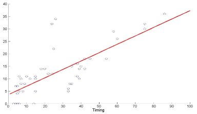
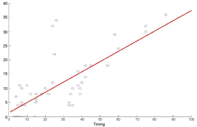
As we told before, 54.28% of patients have been treated in ultra-early surgery, 33.33% in early surgery, and 12.28% in delayed surgery. We investigate how this parameter could influence the outcome in patient's object of our study. In patients in the first group (ultra-early surgery) the outcome was favourable to one year in 32 patients (56.1%), not favourable in 25 patients (43.8%). In patients in the second group (early surgery) the outcome was favourable in 11 patients (34.3%), not favourable in 24 patients (65.7%). In patients in the third group (delayed surgery), the outcome was favourable in 3 patients (23.1%), not favourable in 10 patients (76.9%). We also estimated the correlation (ρ) (values between -1 and 1) between the "surgical timing" value and the three-month and one-year outcome, obtaining the following values: 0.41 at three months and 0.57 at one year, both positive. The covariance σ (surgical timing-mRS) at three months and one year was respectively 1.22 and 1.35, both positive to indicate that the two values positively combined. Finally, through a Linear Regression Model (OLS), we estimated the regression coefficient of the "surgical timing" variable against the mRS (and therefore the outcome), obtaining the following results: 0.53 at three months and 0.62 at one year. This indicates that an increase of one hour between the onset of symptoms and the surgery involves an expected increase in the score in the mRS scale of 0.53 to 3 months and 0.62 in a year. See Figure 3 and Figure 4. To investigate the effect and weight of these two parameters on outcome, we estimated the marginal effects on the outcome of the ICH score (severity of hemorrhage) and surgical timing using the aforementioned linear regression model. We then calculated the regression coefficient of both variables against the mRS obtaining the following results: 0.45 for the ICH score and 0.32 for surgical timing at the three-month; 0.29 for the ICH score and 0.63 for surgical timing at one-year. This means that, although in all cases the coefficients are positive and useful to predict the outcome of the patient, at three months the mRS (early outcome) was more influenced by the score and hence the severity of the ICH. While at 1 year the situation was reversed: The outcome was more affected by surgical timing.
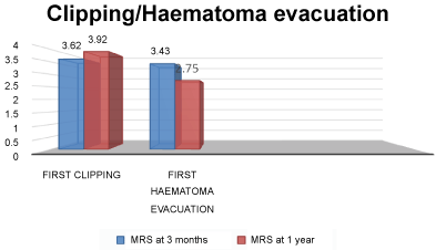
Aneurysm clipping and hematoma evacuation
In our series, 24 of patients with ICH (42.1%) firstly have been submitted to clipping of the aneurysm and then to hematoma evacuation. In 33 patients (57.9%) firstly have been performed hematoma evacuation and then clipping of the aneurysm. In the 48 patients without ICH, only the clipping of the aneurysm was carried out. Figure 5 shows instead the average mRS and therefore the average 3 months and one-year outcome respectively in patients who were first subjected to clipping of the aneurysm or before hematoma evacuation. The best outcome was found in patients who first went to evacuation of hematoma and therefore to the clipping of the aneurysm. However, this analysis was not statistically significant (p-value: 0.1).
Discussion
MCAAs features
The MCA is the largest of the cerebral arteries; its diameter at the origin is about 4 mm (range, 2.4-4.6 mm) and its course begins at the ICA bifurcation and proceeds laterally below the anterior perforated substance within the sylvian fissure dividing into its main trunks. The MCA branches reach the cortical surface of frontal, parietal and temporal opercula lobes. Aneurysms of MCA artery (MCAAs) represent 18-40% of all intracerebral aneurysms and the most frequent location is at the main MCA bifurcation. Because of the variety of MCAAs morphology, size, location and projection, in literature some MCAAs classifications have been proposed in order to identify the best management strategy. Dashi R, et al. [8] proposed a five-types classification of MCA Bifurcation Aneurysms (MCAbifAs), composed by intertruncal MCAbifAs (type 1), inferior MCAbifAs (type 2), lateral MCAbifAs (type 3), insular MCAbifAs (type 4) and complex MCAbifAs (type 5). For each of these types the Author described a different head positioning, Sylvian scissure's dissection and temporary and final clipping strategy. MCAbifAs present more than other kinds of MCAAs a heterogeneous morphology due to their localization on the principal artery. Our study includes all MCAAs that have been considered within three principal groups: Proximal, main bifurcation and distal: MCAAs can be located in the proximal segment of the principal artery (M1) or at the main MCA bifurcation (MCAbif) or distally (M2-M3). Regarding the size, we have distinguished MCAAs in small (< 7 mm), medium (7-14 mm), large (15-24 mm) or giant (> 24 mm); morphologically MCAAs can be saccular, blister-like or fusiform.
Treatment choice
The discussion about the best treatment choice between endovascular coiling/stenting or surgical clipping has just begun years ago. Bracard, et al. [9] evaluated clinical outcome in a series of 140 MCAAs treated by endovascular coiling and reported a persistent occlusion rate of 83.3% at 1 year post-treatment. Brinjikji, et al. [10] confirmed an obliteration rate at follow-up of 82.4% with an overall coiling procedure-related morbidity and mortality of 6% for ruptured aneurysms. Whereas Kim KH, et al. [6] reported an obliteration rate of 93% within the endovascular coiling group of 30 patients, compared with an obliteration rate of 92% in the surgical clipping group of 78 patients. The Author preferred the surgical clipping to the endovascular coiling embolization, and suggested to consider coiling for patients with serious medical problems or where there is the risk of damaging perforating lenticulostriate arteries on the MCA during surgery. During the last years the rapid evolving of endovascular coiling technique has given more experience among neurovascular interventional radiologists and neurovascular surgeons, indeed comparisons in terms of morbidity and mortality between the two techniques result similar. Diaz OM, et al. [11] reported a higher overall rate of complications and retreatment in the endovascular group than surgical one, despite what the functional outcome of the two groups resulted similar at the follow-up. The International Subarachnoid Aneurysm Trial (ISAT) Collaborative Group enrolled 2143 patients with ruptured intracranial aneurysms, of which patients with middle cerebral artery aneurysms are under-represented because the anatomy of the aneurysmal neck is more often unfavourable for endovascular treatment [12]. Microsurgical clipping is still the preferred treatment modality in most centres due to the relative superficial position of these aneurysms and to the ICH often associated: The surgical approach gives a straightforward trajectory to the aneurysmal dome. Moreover surgery allows releasing Cerebrospinal Fluid (CSF) which improves management of the increased intracranial pressure.
Outcome
Our study targets ruptured MCAAs surgically treated and their outcome in relation with clinical evaluation, surgery timing, topographical features of MCAAs and the high percentage of an intra-cerebral haematoma (ICH = 54.3%). We have included in our study only surgically treated patients with ruptured MCAAs: There is a prevalence of MCAbifAs, which is the most superficial line of the MCA (67%). Most of patients have been treated within 12 hours from the symptoms onset (54.3%) and in these patients we have reached the best results in terms of outcome, evaluated with the Modified Rankin Scale (mRS). Furthermore these patients show a re-bleeding rate of 8.7%, the lowest within the three groups (ultra-early, early and delayed surgery). There is a positive relationship between the surgical timing and mRS Score, meaning that an increase of one hour between the onset of symptoms and surgery causes a worsening in 3 months and 1 year outcome. Therefore all patients should be treated within 12 hours from the onset of symptoms, in order to improve outcome. ICH is an independent and relevant parameter to evaluate in terms of choice for timing and treatment modality [13-15]. Our analysis proves that an increase of 1 point of ICH score has a linear relationship with an increase in the mRS scale as to 3 months as to 1 year outcome. The early outcome is more influenced by ICH severity score, while at 1 year is more influenced by surgical timing. Hence, the higher morbidity and mortality are strictly related to the brain swelling due to the ICH volume. A forward hematoma evacuation allows earlier cisternal decompression in order to reach more easily and quickly aneurysmal dome. In our series, patients who went to first haematoma evacuation and then aneurysmal clipping show a better outcome than other.
Limits
This study has several limits. First, this is a retrospective study without randomization, and there was not an endovascular control group for comparison of the procedure-related outcome. Moreover the small number of patients in our series doesn't assure the best statistical significance.
Conclusion
Concerning result of our analysis, in MCAAs patients, the presence of ICH strongly affects the outcome with a marked increase in mortality and morbidity. Surgical timing significantly influences the outcomes and in patients with a poor-grade entry and with Intraparenchymal bleeding, ultra-early surgery should always be taken into account.
Conflicts of Interest
The authors declare that they have no conflicts of interest.
References
-
Hallout S (2015) Surgical Treatment of Middle Cerebral Artery Aneurysms without Using Indocyanine Green Video angiography Assistance: Retrospective Monocentric Study of 263 Clipped Aneurysms. World Neurosurg 84: 972-977.
-
Baharoglu MI, Lauric A, Safain MG, et al. (2014) Widening and High Inclination of the Middle Cerebral Artery Bifurcation Are Associated With Presence of Aneurysms. Stroke 45: 2649-2655.
-
Morgan MK, Mahattanakul W, Davidson A, et al. (2010) Outcome for middle cerebral artery aneurysm surgery. Neurosurgery 67: 755-761.
-
Backes D, Rinkel GJ, Laban KG, et al. (2016) Patient- and Aneurysm-Specific Risk Factors for Intracranial Aneurysm Growth: A Systematic Review and Meta-Analysis. Stroke 47: 951-957.
-
Orz Y, AlYamany M (2015) The impact of size and location on rupture of intracranial aneurysms. Asian J Neurosurg 10: 26-31.
-
Kim KH, Cha KC, Kim JS, et al. (2013) Endovascular coiling of middle cerebral artery aneurysms as an alternative to surgical clipping. J Clin Neurosci 20: 520-522.
-
Wang HJ, Ye YF, Shen Y, et al. (2014) Surgical Treatment of Poor Grade Middle Cerebral Artery Aneurysms Associated with Large Sylvian Hematomas Following Prophylactic Hinged Craniectomy. J Huazhong Univ Sci Technolog Med Sci 34: 716-721.
-
Dashti R, Hernesniemi J, Niemel M, et al. (2007) Microneurosurgical management of middle cerebral artery bifurcation aneurysms. Surg Neurol 67: 441-456.
-
Bracard S, Abdel-Kerim A, Thuillier L, et al. (2010) Endovascular coil occlusion of 152 middle cerebral artery aneurysms: initial and midterm angiographic and clinical results. J Neurosurg 112: 703-708.
-
Brinjikji W, Lanzino G, Cloft HJ, et al. (2011) Endovascular Treatment of Middle Cerebral Artery Aneurysms: A Systematic Review and Single-Center Series. Neurosurgery 68: 397-402.
-
Diaz OM, Rangel-Castilla L, Barber S, et al. (2012) Cerebrovascular Middle Cerebral Artery Aneurysms : A Single-Center Series Comparing Endovascular and Surgical Treatment. World Neurosurg 8: 322-329.
-
Molyneux A, Kerr R, Stratton I, et al. (2002) International Subarachnoid Aneurysm Trial (ISAT) of neurosurgical clipping versus endovascular coiling in 2143 patients with ruptured intracranial aneurysms : a randomised trial. Lancet 360: 1267-1274.
-
Stapleton CJ, Walcott BP, Fusco MR, et al. (2015) Surgical management of ruptured middle cerebral artery aneurysms with large Intraparenchymal or sylvian fissure hematomas. Neurosurgery 76: 258-264.
-
Jabbarli R, Reinhard M, Roelz R, et al. (2016) Intracerebral Hematoma Due to Aneurysm Rupture: Are There Risk Factors Beyond Aneurysm Location? Neurosurgery 78: 813-820.
-
Fukuda H, Hayashi K, Moriya T, et al. (2015) Intrasylvian hematoma caused by ruptured middle cerebral artery aneurysms predicts recovery from poor-grade subarachnoid hemorrhage. J Neurosurg 123: 686-692.
Corresponding Author
Benedetta Fazzolari, MD, Department of Neurology and Psychiatry, Neurosurgery Unit, Sapienza University of Rome, Viale del Policlinico, 155, 00100, Rome, Italy.
Copyright
© 2017 Ruggeri AG, et al. This is an open-access article distributed under the terms of the Creative Commons Attribution License, which permits unrestricted use, distribution, and reproduction in any medium, provided the original author and source are credited.