'Adenomyosis': The Enigma
Adenomyosis was classically defined by Bird, et al. as "Benign invasion of endometrium in the myometrium producing a diffusely enlarged uterus which microscopically exhibits ectopic nonneoplastic endometrial glands and stroma surrounded by hypertrophic and hyperplastic myometrium" [1]. It is this hypertrophic and hyperplastic myometrium that needs to be tailored in the surgical management of adenomyosis and hence holds a lot of importance in the entire discussion of adenomyosis.
Etiology
The exact etiology of adenomyosis is not known hence atleast 4 theories have been postulated [2].
1. The first and most popular theory is that adenomyosis develops from the invagination of endometrium into the myometrium. Steroid hormone studies have shown that adenomyotic tissue exhibits higher expression of estradiol receptors than does eutopic endometrium. The increased response to estrogen may facilitate the overall invagination of endometrium.
2. A second theory is that adenomyosis develops de novo from embryologically misplaced pluripotent mullerian remnants. Extra-uterine sites for adenomyosis such as that in the rectovaginal septum supports this theory.
3. Theory of intralymphatic intramyometrial spread.
4. Bone marrow stem cell origin of adenomyosis.
Classification
The surgical/histological classification of adenomyosis was given by Grimbiz, et al. in 2013 [3] as shown in Table 1.
Diagnosis
Clinically the patient may present with any or all of the below mentioned symptoms like abnormal uterine bleeding, dysmenorrhea, chronic pelvic pain, dyspareunia, poor reproductive outcomes (infertility, or recurrent miscarriages).
Imaging
Ultrasound
A Transvaginal ultrasound forms a very important tool in the diagnosis of adenomyosis with the suggestive signs of an asymmetrically and diffusely enlarged uterus with increased myometrial echogenicity or linear hyperechoic bands extending deep into the myomectrium and the presence of myometrial cysts as shown in Figure 1.
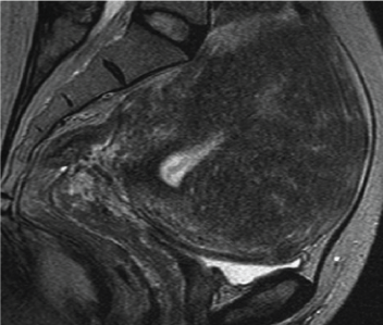
MRI
On T2 weighted images a large asymmetrically enlarged uterus along with myometrial heterogeneity, and presence of myometrial cysts along with enlargement of the junctional zone (the innermost myometrial layer) can be seen in adenomyosis [4]. MRI has very high specificity and positive predictive value of more than 90 percent and is especially useful if fibroids coexist in the same patient to differentiate between the two (Figure 2).
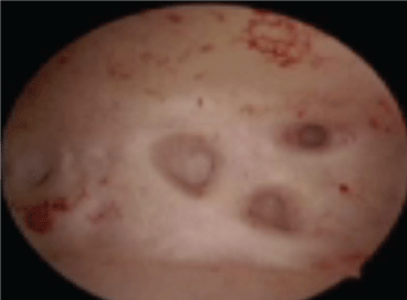
Hysteroscopy
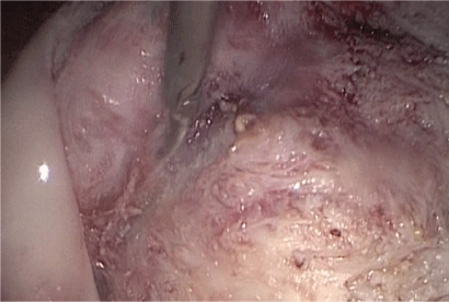
In adenomyosis irregular endometrium with pitting endometrial defects altered vascularization and cystic hemorrhagic lesions maybe observed on hysteroscopy (Figure 3).
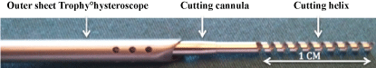
A Gordt's spirotome may be used to get a hysteroscopic biopsy from the suspicious areas (Figure 4).
Treatment
The basic principle of medical management is by means of hormonal treatment which only work as agents for 'Suppression of the Disease' whereas Surgery which involves excision of the lesions, remains the mainstay for the treatment of the disease [5,6]. Hence continuous oral contraceptive pills, Depo-Provera, Levonorgestrel IUCD, Danazol, IUCD and GnRH agonists all work as temporary methods for suppression of the disease.
Alternative methods
Treatments such as High Frequency Ultrasound (HIFU) and Uterine Artery Embolization (UAE) have been proposed for treatment of selective cases of adenomyosis [5,7]. Their role is still controversial and data on pregnancy outcome is scanty. Therefore presently alternate treatments should be proposed in women with no desire for future pregnancy [8]. Also UAE for adenomyosis is technically different from when it is performed for fibroids as at angiography adenomyosis shows a reduction in arterial pattern but increase in microvessel density making the use of polyvinyl alcohol particles less effective. Hence the absence of a good embolic agent suited to adenomyosis currently makes this procedure less effective than when it is used for fibroids.
Fertility Sparing Surgeries
As women with adenomyosis suffer not only from an increased incidence of infertility but also from an increased incidence of recurrent miscarriages, along with menorrhagia and dysmenorrhea, women desirous of future pregnancy have been treated by Extensive Radical Adenomyomectomies for symptom relief and to achieve a good pregnancy outcome [9]. With Extensive radical adenomyomectomies the resultant scars were not strong enough to withstand subsequent pregnancy hence several surgeons like Osada came up with special techniques like Triple Flap technique where after a radical adenomyomectomy the myometrium is sutured back as overlapping flaps to ensure stronger scars that can withstand subsequent pregnancy. This procedure results in more than 70 percent spontaneous conception rate. Also along with good reproductive outcome women get a lot of relief from the dysmenorrhea and menorrhagia. We advise that subsequent pregnancies should be delivered by elective caesarean section only.
The Sunrise Laparoscopic Flap Adenomyomectomy
The Procedure;
The primary trocar is inserted at the modified Palmers point and then 3 secondary trocars are 1) placed under vision lateral to the inferior epigastric vessels on each side and one suprapubic.
2) Dilute Vasopressin is then infiltrated into the uterus (40 units in 200 ml of normal saline).
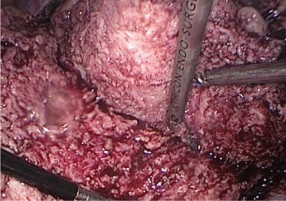
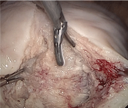
3) A vertical incision is then given over the uterus up to the endometrial cavity, 1 cms of margin of myometrium is left over the basal is layer of endometrium (Figure 5).
4) On the serosal layer side 1 cms of margin of myometrium is left inside by tunneling. All the in between myometrium is then excised (Figure 6).
5) The end result is that the uterus is left with 1 cm myometrium over the endometrium and another 1 cm myometrium under the serosa (Figure 7).
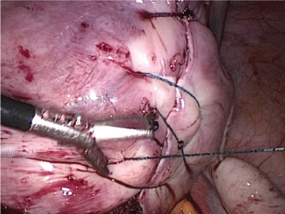
6) To make this remaining myometrium withstand subsequent pregnancy the uterus is then sutured with V-lock continuous sutures as reinforcing flaps.
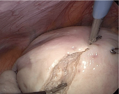
Technical challenges of this procedure are that compared to myomas there are no distinct planes/capsule hence the excision of tissue is entirely arbitrary, left to the discretion of the surgeon. Also the adenomyomatous tissue is difficult to hold and manipulate compared to normal myometrium. Secondly the surgeon has to have good experience with laparoscopic suturing as extensive flap suturing is mandatory for the success of this procedure (Figure 8). Also the surgeon has to be good at lateral pelvic wall dissection laparoscopically as uterine artery clipping at origin maybe required in some cases for adequate hemostasis. All these challenges make this procedure a very advanced laparoscopic procedure to be performed by experts only.
Laparoscopic Endomyometrectomy [Gross Wedging of the Uterus]
This procedure is designed for women who are not desirous of future pregnancy and are between 35 and 45 yrs of age. The principle of this procedure is that the disease parts namely the endometrium and myometrium is removed with conservation of the no diseased part of the uterus. As a large proportion of the blood supply of the ovary is derived from the uterine artery (almost 50-60%), we aim at conserving this blood supply by performing a laparoscopic endomyometrectomy in young patients who are not desirous of having more children and yet wish to retain their uterus hence giving them an additional advantage of continued hormonal function via the untouched ovarian and uterine blood supply.
Step by step explanation of the technique
1. General anesthesia given in supine position.
2. Urinary bladder is emptied and primary trocar inserted via direct closed entry technique at modified palmar's point (an arbitrary point medial to palmar's point and lateral to Lee Huang point in the upper quadrant of abdomen).
3. Pneumoperitoneum created and three secondary trocars inserted under vision, two lateral trocars on both sides (right and left) and one central trocar.
4. Diluted vasopressin (40 units vasopressin in 200 ml saline) is injected in the uterus (Figure 9).
5. Uterus is manipulated using myoma screw from right port.
6. Uterine incision is given 1 cm medial to the cornual structures bilaterally using ultrasonic energy source.
7. This line of incision is followed caudally keeping a margin of 1 cm from the lateral uterine wall which has uterine vessels in its course.
8. Incision is continued till the level of uterovesical fold anteriorly and just above the uterine attachment of uterosacral ligaments posteriorly.
9. At this level lateral incisions are merged and entire myometrial tissue with endometrial cavity (endomyometrium) is excised.
10. Bilateral uterine walls are sutured and approximated with each other and cervix.
11. Excised endomyometrium is delivered out using power morcellator.
The end result is very small size uterus which is actually non-functional. Patient should be advised that she would not have her menses, slight cyclical spotting is possible though.
Advantages
1. Uterine vessels are left untouched, hence ovarian blood supply is maintained, hence no hormonal disturbance.
2. All uterine ligaments are intact, uterosacral ligament and pericervical ring stays intact, hence less chances of prolapse.
3. Psychological relief for the patient that her uterus is still preserved and only diseased part is taken out.
References
- Bird CC, McElin TW, Manalo-Estrella P (1972) The elusive adenomyosis of the uterus revisited. Am J Obstet Gynecol 112: 583-593.
- Garcia L, Isaacson K (2011) Adenomyosis: review of the literature. J Minim Invasive Gynecol 18: 428-437.
- Grimbiz (2013) Classification of Adenomyosis. Fertility Sterility.
- Mayumi Takeuchi, Kenji Matsuzaki (2011) Adenomyosis: Usual and unusual imaging manifestations, pitfalls, and problem-solving MR imaging techniques. Radiographics 31: 99-115.
- Interventional procedure guidance (2013) Uterine artery embolization for treating adenomyosis.
- Pereira RMA, Zanatta A, PHM Bianchi, et al. (2009) Laparoscopic Adenomyosis resection for symptomatic infertile patients: Case Series. J Minim Invasive Gynecol 16: S117.
- Huann Cheng Horng, Ching Hui Chen, Chih Yao Chen, et al. (2014) Uterine Sparing Surgery for Adenomyosis. Taiwan J Obstet Gynaecol 53: 3-7.
- Alabiso G, Alio L, Arena S, et al. (2016) Adenomyosis: What the patient needs. J Minim Invasive Gynecol 23: 476-488.
- Osada H, Silber S, Kakinuma T, et al. (2011) Surgical procedure to conserve the uterus for future pregnancy in patients suffering from massive adenomyosis. Reprod Biomed Online 22: 94-99.
Corresponding Author
Dr. Nikita Trehan, Sunrise Hospitals, India; International Modern Hospital, Dubai, United Arab Emirates, Tel: +919811000070.
Copyright
© 2017 Rahman H, et al. This is an open-access article distributed under the terms of the Creative Commons Attribution License, which permits unrestricted use, distribution, and reproduction in any medium, provided the original author and source are credited.