How to Evacuate Surgical Smoke While Using Laparoscopic Coagulation Shears
Abstract
Introduction
During laparoscopic surgery, the production of smoke by energized dissecting devices is a problem because it can obstruct the view of the operative field. The products of combustion may also contain hazardous chemicals, such as benzene, toluene, viable cancer cells or viruses. However, these devices, in particular Laparoscopic Coagulation Shears (LCS), are often essential during laparoscopic surgery. We suggest a simple evacuation system to resolve this problem using intravenous tubing.
Material and surgical technique
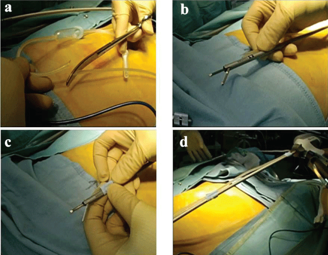
The intravenous tubing is cut diagonally, proximal to the drip chamber and sterile spike. The cut end of the tubing is taped to the shaft near the active blade side on the LCS. The luer-lock side of the tubing is connected to a suction source. The smoke and vapor arising at the dissection point during the use of the LCS are removed by the application of continuous suction to the tubing.
Discussion
We describe a method for the efficient evacuation of combustion products generated by the intraoperative use of the LCS using an inexpensive and easily available device.
Keywords
Laparoscopic coagulation shears, Laparoscopic surgery, Surgical smoke
Introduction
Studies have shown that the substances produced by electro-coagulation devices, such as Laparoscopic Coagulation Shears (LCS), contain hazardous substances, including cellular material, tumor cells [1,2], viruses [3], and chemical substances (benzene, toluene, etc.) [4,5]. Tomita, et al. reported that the amount of smoke from electro-coagulation that condensed on 1 g of mucosal tissue of a canine tongue was equivalent to that from three to six cigarettes, with respect to total mutagenicity [6]. The Japanese medical society has recommended that operating room personnel and surgeons avoid inhaling or being exposed to smoke containing harmful chemical substances [7,8]. During laparoscopic surgery, smoke is evacuated from the laparoscopic port using a smoke extraction system. However, this system can lead to diffusion of mist or vapor in the intra-abdominal space between the site of energy application and the laparoscopic suction port. Diffusion and delayed evacuation of the mist or vapor is a constant problem for surgeons performing laparoscopic surgery.
We suggest an innovative measure to evacuate the mist or vapor at the point of energy application using an easily available and inexpensive device. This novel system provides clearer vision and safety for staff in the operative room.
Materials and Surgical Technique
Readily available materials, including surgical tape, intravenous infusion tubing, and the LCS are used to make the smoke evacuation system. The Sonicision™ cordless system (Medtronic Company, Minneapolis MN USA) and a Terufusion Volumetric Solution Administration Set (Terumo Company, Japan) were used. Figure 1a shows the intravenous tubing cut diagonally at the proximal side of the drip chamber and the sterile spike. The cut end is fixed with surgical tape at the active blade side on the shaft, about 3 cm from the active blade (Figure 1b and Figure 1c). The tubing is fixed at three sites son the LCS shaft with surgical tape, but the tubing is not fixed to the LCS generator (Figure 1d), because rotation is not possible if the tubing is fixed to it. The luer-lock side is connected directly to the suction source. Intra abdominal pressure is usually set to 10 cm H2O in high-flow insufflation mode (20~45 L/min). When using the LCS for dissection, the combustion byproducts are evacuated continuously via the tubing to the suction device, and suction pressure is controlled with the usual regulator. With continuous smoke suction, the intra abdominal pressure is normally reduced by around 7-8 cm H2O. However, the reduction of the intra abdominal pressure rarely obstructs the field of vision. The LCS dissector with the tubing attached is easily introduced into the intra peritoneal space through a 12 mm, but not a 5 mm, laparoscopic port.
Discussion
The intraoperative use of energy devices results in safe dissection and excellent hemostasis, which has fostered the global spread of laparoscopic surgery. This technology, in particular LCS, is virtually essential in laparoscopic surgery. There is evidence that operating room staff could be exposed to surgical smoke containing hazardous chemicals, such as benzene, toluene, ethanol, 1,2-dichloroethane, ethylbenzene, and styrene [4,5] and even viable malignant cells [1,2]. Thus, operating room staff is advised to avoid or minimize their exposure to surgical smoke. Furthermore, absorption of these chemicals across the serosal surfaces is possible, and minor elevations in carboxyhemoglobin levels resulting from the intra-abdominal use of electro cautery have been demonstrated in a laparoscopic porcine model [9]. In, et al. reported that viable cancer cells contained in surgical smoke from ultrasonic scalpels grew at 16 of 40 injection sites after implantation in mice [2]. These results suggest that patients undergoing laparoscopic surgery are at increased risk of absorption of hazardous chemicals in the body and could develop local recurrence after surgery for advanced cancers. Several studies have shown that viable cancer cells in surgical smoke could be responsible for port site metastasis [10,11]. The internal lumen of used tubing could also contain viable cancer cells, leading to the potential for local recurrence in surgical fields.
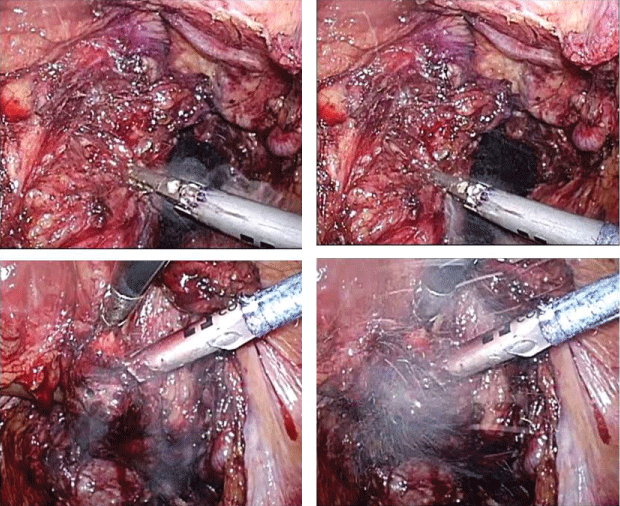
The medical societies in Japan have recommended the control of surgical smoke by local exhaust [7]. However, most surgeons and perioperative nurses in Japan remain indifferent to this issue. Thus, the operating room staff can be exposed to harmful chemicals when the pneumoperitoneum is released from the laparoscopic port after LCS dissection, or at the end of laparoscopic procedures. Conventional port site suction systems can lead to diffusion of a mist from the site of dissection throughout the intraperitoneal space. The scope assistant, surgeon and surgical assistant often experience stress due to the repeated movement of the scope, in order to prevent fogging of the lens during LCS dissection. The smoke evacuation system described herein removes the mist at the actual site of production during LCS dissection (Figure 2). The lens of the laparoscope is rarely obscured using this system.
Several issues were observed when using this system. Firstly, it took five minutes to prepare the system before surgery. Secondly, the LCS dissector with the tubing attached could not be introduced into the intra-abdominal space through a 5 mm port. Thirdly, the tubing occasionally interfered with the surgeon's handling of the LCS dissector. Several studies have proposed a technical solution to efficiently remove surgical smoke via a conventional port [12,13]. However, our suggested system could decrease the risk of exposure to harmful chemicals and provide a clear view by the continuous evacuation of mist at the site of origin. Ideally, the LCS dissector could be re-designed in the future with an improved smoke evacuation system, which could be introduced through a 5 mm laparoscopic port.
In conclusion, this smoke evacuation system provides surgeons and operating room staff with a safe environment, improves the intraoperative view of the operating site, and reduces stress. We believe that these experiences suggest the need to develop a new LCS device with a smoke evacuation system that most surgeons would desire for laparoscopic surgery.
Acknowledgements Section
All authors have declared that they have no conflicts of interest or any financial disclosures.
References
- Fletcher JN, Mew D, DesCôteaux JG (1999) Dissemination of melanoma cells within electrocautery plume. Am J Surg 178: 57-59.
- In SM, Park DY, Sohn IK, et al. (2015) Experimental study of the potential hazards of surgical smoke from powered instruments. Br J Surg 102: 1581-1586.
- Garden JM, O'Banion MK, Shelnitz LS, et al. (1988) Papillomavirus in the vapor of carbon dioxide laser-treated verrucae. JAMA 259: 1199-1202.
- Choi SH, Kwon TG, Chung SK, et al. (2014) Surgical smoke may be a biohazard to surgeons performing laparoscopic surgery. Surg Endosc 28: 2374-2380.
- Fitzgerald JE, Malik M, Ahmed I (2012) A single-blind controlled study of electrocautery and ultrasonic scalpel smoke plumes in laparoscopic surgery. Surg Endosc 26: 337-342.
- Tomita Y, Mihashi S, Nagata K, et al. (1981) Mutagenicity of smoke condensates induced by CO2-laser irradiation and electrocauterization. Mutat Res 89: 145-149.
- Kubo H (2013) Medical engineering, electrical instruments and medical gas, guideline for surgical practice (in Japanese). Nihon ShujutsuIgaku Kaishi 34: 98-113.
- Barrett WL, Garber SM (2003) Surgical smoke: a review of the literature. Is this just a lot of hot air? Surg Endosc 17: 979-987.
- Wu JS, Monk T, Luttmann DR, et al. (1998) Production and systemic absorption of toxic byproducts of tissue combustion during laparoscopic cholecystectomy. J Gastrointest Surg 2: 399-405.
- Cavina E, Goletti O, Molea N, et al. (1998) Trocar site tumor recurrences. May pneumoperitoneum be responsible? Surg Endosc 12: 1294-1296.
- Kazemier G, Bonjer HJ, Berends FJ, et al. (1995) Port site metastases after laparoscopic colorectal surgery for cure of malignancy. Br J Surg 82: 1141-1142.
- Takahashi H, Yamasaki M, Hirota M, et al. (2013) Automatic smoke evacuation in laparoscopic surgery: a simplified method for objective evaluation. Surg Endosc 27: 2980-2987.
- Mattes D, Silajdzic E, Mayer M, et al. (2010) Surgical smoke management for minimally invasive (micro) endoscopy: an experimental study. Surg Endosc 24: 2492-2501.
Corresponding Author
Mitsuaki Morimoto, MD, Department of Gastrointestinal Surgery, Jichi Medical University, 3311-1 Yakushiji, Shimotsuke, Tochigi 329-0498, Japan, Tel: +81-285-5-7371, Fax: +81-285-44-3234.
Copyright
© 2017 Morimoto M, et al. This is an open-access article distributed under the terms of the Creative Commons Attribution License, which permits unrestricted use, distribution, and reproduction in any medium, provided the original author and source are credited.


