Sudden Vision Loss: More than Just an Ophthalmic Diagnosis! A Case Report
Abstract
Introduction: Tuberculosis is a public health concern worldwide with India being the highest (26%). However, tuberculosis of paranasal sinuses was unusual even in countries with a high prevalence of the disease. Isolated involvement of sphenoid sinus by disease is a rare entity.
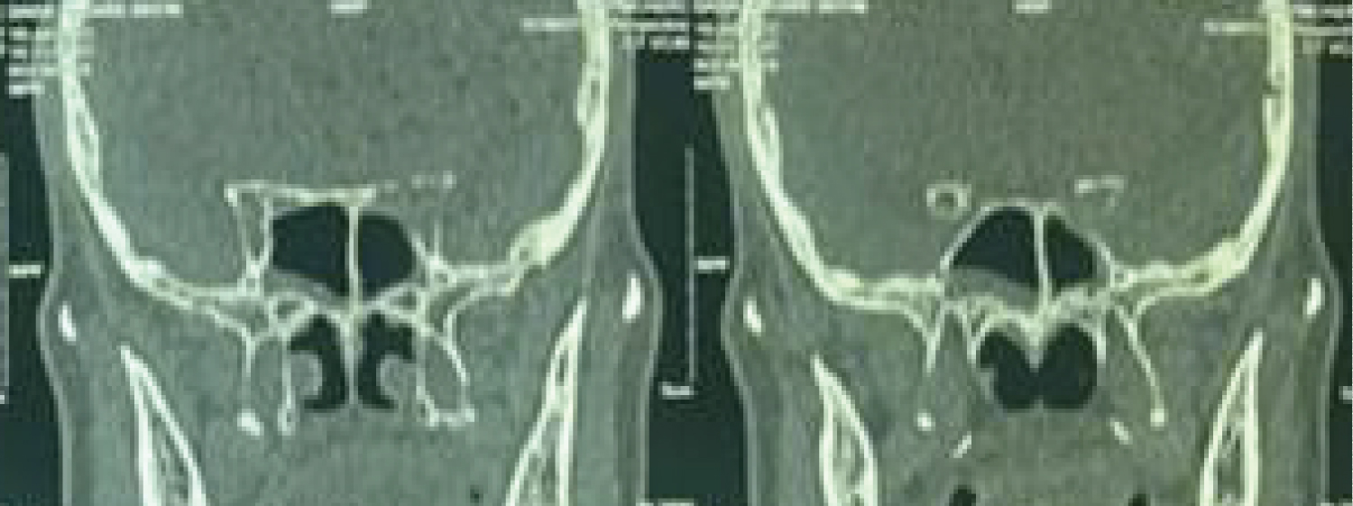
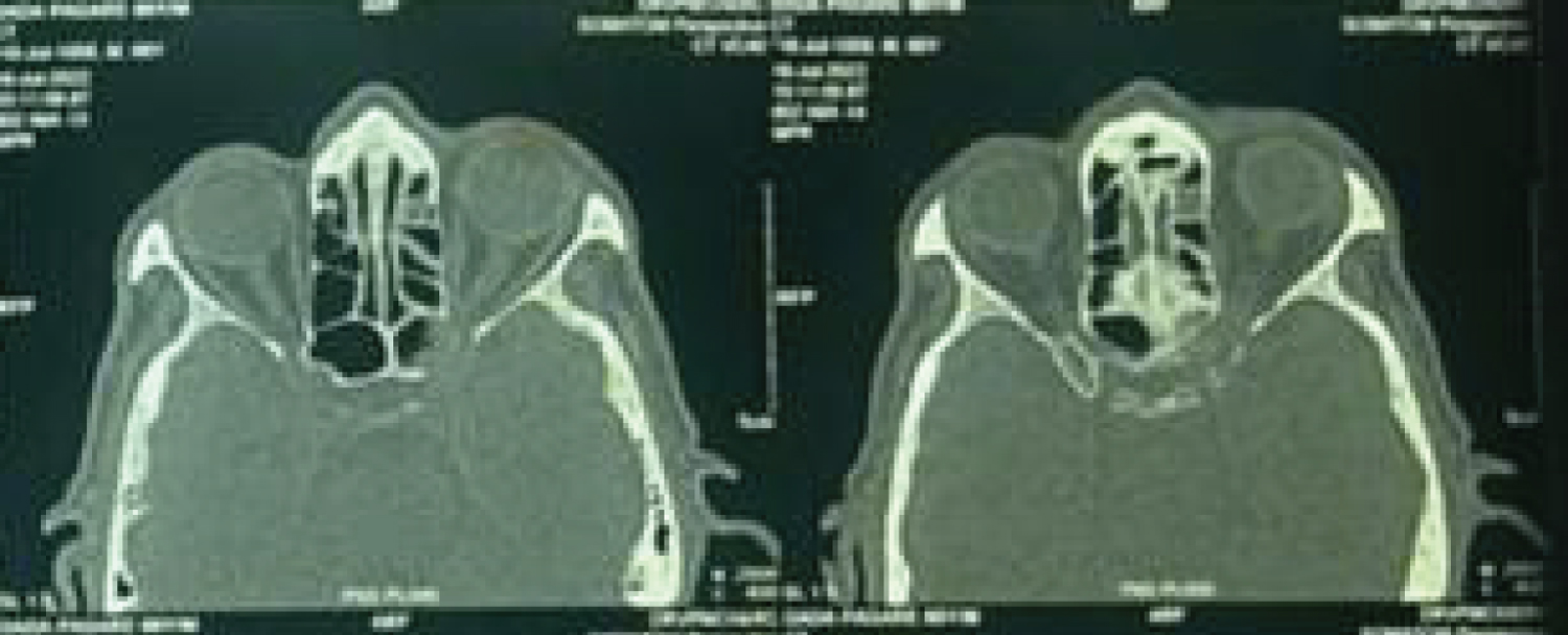
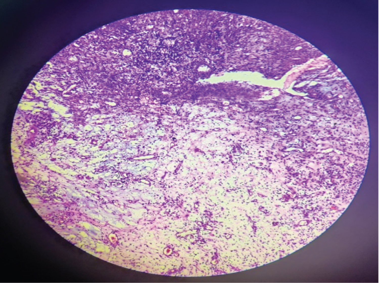
Case report: A 66-year-old male patient presented with the chief complaints of sudden diminution of vision in the left eye. The left eye vision was finger counting at 3 meters and that of the right eye was normal. Non-Contrast Computed Tomography of the nose and paranasal sinuses showed an ill-defined soft tissue density in the left sphenoid sinus abutting the left optic nerve with erosion of superior wall of left orbit. The lesion was removed completely by Transnasal Endoscopic Approach. The left optic nerve decompression was also done simultaneously. There was a significant improvement in the vision immediately postoperatively. Histopathology report showed epitheloid cell granulomas with giant cells suggestive of tuberculosis.
Discussion: As tuberculosis is a systemic disease, it can affect any part of body. In our case, it mainly affects vision of the patient without any other complaints. It has to differentiate mainly from malignancy when old age patients are affected. Once diagnosed tuberculosis responds well to Anti Tubercular Treatment (ATT).
Conclusion: We should consider tuberculosis as a first differential diagnosis in cases of isolated sphenoid sinus lesions mainly in developing countries. Optic nerve decompression should be considered in those with affected vision.
Keywords
Tuberculosis, Sphenoid sinus, Vision, Computed tomography, Endoscopic
Introduction
Tuberculosis is a public health concern worldwide with India being the highest (26%).In 2017, the World Health Organization (WHO) gave an estimated incidence of 1339 million cases in India. However, tuberculosis of paranasal sinuses was unusual even in countries with a high prevalence of the disease. Isolated involvement of sphenoid sinus by disease is a rare entity. Isolated sphenoid sinus lesions account for only 2.7 to 3% of the pathologies of the paranasal sinuses; common etiologies are inflammatory polyps, fibro-osseous lesions, neoplastic mass, and fungal sinusitis [1,2]. We encountered one such unusual case of Tuberculosis presented as an isolated sphenoid sinus lesion with vision loss.
Case Report
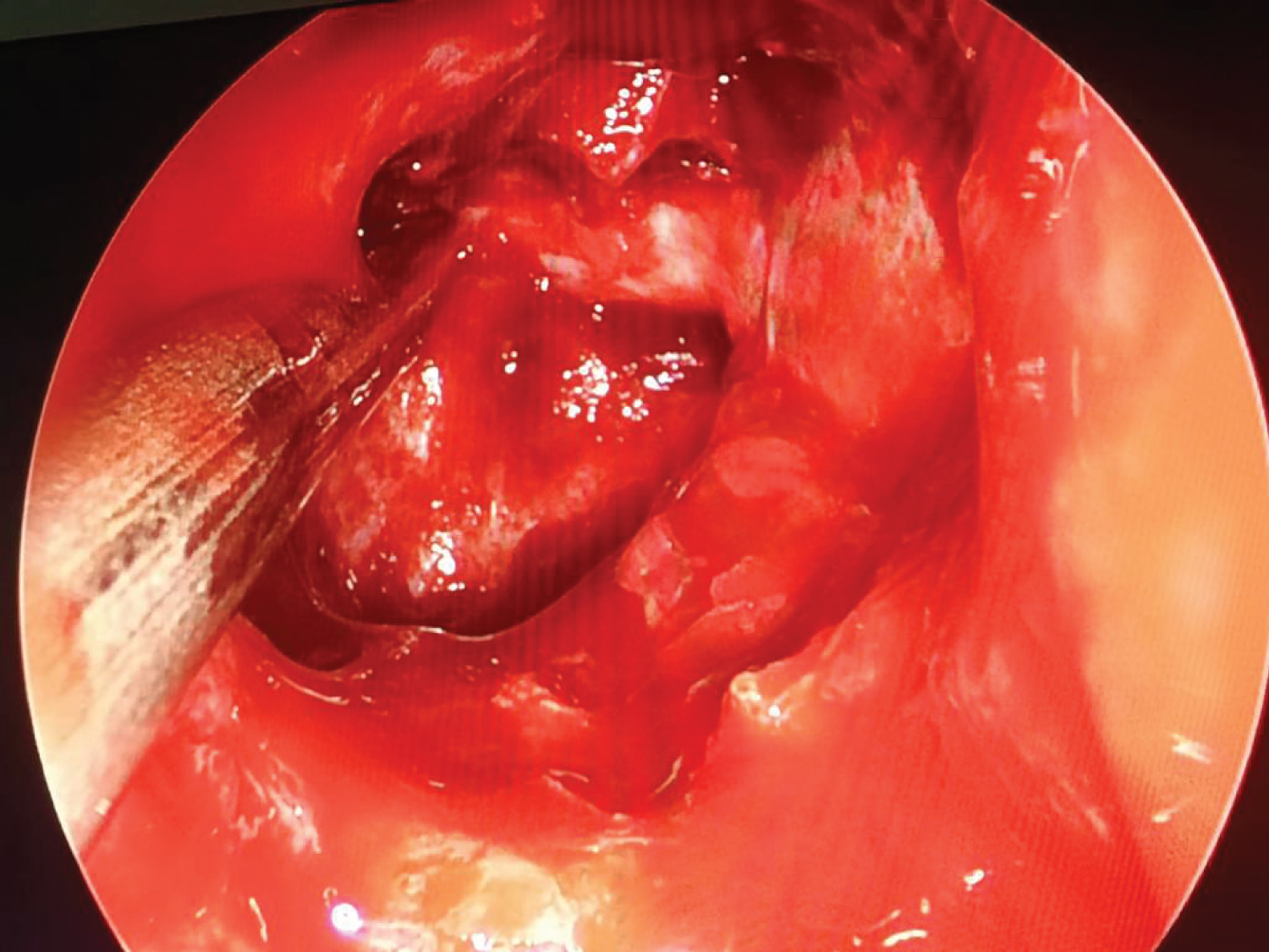
A 66-year-old male patient presented with the chief complaints of sudden diminution of vision in the left eye since 10 days. There was no history of nasal blockage, nasal discharge, facial swelling, facial pain, or eye pain. On Anterior Rhinoscopy there was deviated nasal septum towards left. Paranasal sinus tenderness was not present. Rest ear, nose and throat examination unremarkable. Vision assessment done pre-operatively which was 6/12 for right eye and finger counting at 3 meters for left eye. On examination of neck there was no palpable lymphadenopathy. We have done Non-Contrast Computed Tomography Scan (NCCT) of nose and paranasal sinus which revealed an ill- defined soft tissue density in the sphenoid sinus of left side with left optic canal abutting the superior rectus muscle and left optic nerve with erosion seen in superior wall of left sphenoid (Figure 1 and Figure 2). Patient was taken under general anaesthesia for endoscopic removal of the mass. Intra operatively there was unhealthy pale pinkish gritty mass seen in the left sphenoid sinus which was associated with bony erosion of its superolateral wall. The mass was seen compressing over the left optic nerve till the level of optic chiasma. Complete removal of the mass done and left optic nerve decompression was also done simultaneously to relive pressure on the optic nerve (Figure 3). The mass then sent for histopathological examination. Post-operative vision on day 1 assessment there was significant improvement in left eye. The vision improved till 6/24 from figure counting. There was a significant improvement in left eye vision immediately post-operatively. Histopathological report showed ill formed epitheloid cell granulomas with giant cells surrounded by cuff of lymphocytes suggestive of granulomatous inflammation suggestive of mycobacterium tuberculosis (Figure 4). We could not suspect tuberculosis intraoperatively so we have not sent sample for microbiological examination. By age and radiological findings we thought of malignancy as a first diagnosis but surprisingly it turned out to be Tuberculosis. The patient was kept firstline Anti-Tubercular Therapy (ATT) according to RNTCP regimen for 6 month. On Follow up after a week vision assessment showed improvement on left eye to almost normal i.e. 6/9. The vision assessment on subsequent follow up of 3 months was same i.e. 6/9 for the left eye and the patient was happy and regularly taking ATT.
Discussion
Morgagni in 1761 reported the first case of paranasal sinus tuberculosis [3].Kernan reported the first case of tuberculosis originating in the sphenoid sinus [4]. Bhat, et al. [1] reported two cases of sphenoid sinus tuberculosis in which endoscopic sphenoidotomy was done for both patients and later anti-tubercular therapy was started after confirmation by histopathology report and the vision improvement was assessed on follow-ups. Clinically patients can present with headache (65%) mainly in the region of vertex, nasal obstruction (22%), post nasal drip (11%). The symptoms pertaining to vision loss (15%) and cranial nerve palsies (10%) occur rarely [5]. In our case, patient was mainly having vision loss and there were no other symptoms. Turel MK reported a case of sphenoid sinus tuberculosis presenting with transient diplopia of 3 months duration and gradual loss of vision for 1 month. An endoscopic biopsy was done but there was no vision improvement [6]. In our case, optic nerve decompression was also done which gave immediate vision improvement for the patient. Whenever there is such a case in which there is vision loss along with radiological findings in sinuses then we should also send sample for microbiological examination along with histopathological examination so the main underlying organism can be accurately detected which can lead to accurate treatment. Sharma and Barua reported two cases of sphenoid sinus involvement with extension to the cavernous sinus in one and the posterior ethmoids in the other [7]. In Various studies there was no significant improvement in vision basically in inflammatory lesion but we were lucky to get significant improvement in vision might be because of early intervention and also patient was not having any other comorbidities.
Conclusion
We should consider tuberculosis as first differential diagnosis when a patient presents with decreased vision along with a CT scan findings of soft tissue density in the sphenoid sinus; in a developing country. Optic nerve decompression should be considered in those patients with affected vision.
Conflict of Interest
All authors stated that there is no conflict of interest.
Funding Source
There is no funding source.
Ethical Approval Statement
All authors stated that this publication sent after getting approval from Ethical Commitee of our institute (Dr. Vasantrao Pawar Medical College and Research Centre).
Acknowledgement
There is no specific acknowledgement for this article.
References
- Bhat VS, Samatha KJ, Devika T, et al. (2020) Tuberculosis of Sphenoid Sinus: Report of Two Cases. J Health Allied Sci 10: 42-45.
- WHO (2018) Global Tuberculosis Report 2018. World Health Organization, Geneva.
- Shah NJ, Prashanth V, Garg S, et al. (2010) Primary tuberculosis of the ethmoid and sphenoid sinuses - a rare entity. Bombay Hosp J 52: 86-89.
- Kernan JD (1919) A case of tuberculosis of the sphenoid sinus. Laryngoscope 29: 276-279.
- Kim SW, Kim DW, Kong IG, et al. (2008) Isolated sphenoid sinus diseases: Report of 76 cases. Acta Otolaryngol 128: 455-459.
- Turel MK, Rajshekhar V (2013) Sphenoid sinus tuberculosis: A rare cause of visual dysfunction in an adolescent girl. Neurol India 61: 179-180.
- Sharma SC, Baruah P (2003) Sphenoid sinus tuberculosis in children-a rare entity. Int J Pediatr Otorhinolaryngol 67: 399-401.
Corresponding Author
Dr. Shashikant Anil Pol, Associate Professor, Department of ENT and Head Neck Surgery, Senior Staff Quarters, Dr. Vasantrao Pawar Medical College & Research Centre, Adagaon, Nashik, India, Tel: 7507814025
Copyright
© 2023 Padghan B, et al. This is an open-access article distributed under the terms of the Creative Commons Attribution License, which permits unrestricted use, distribution, and reproduction in any medium, provided the original author and source are credited.