The Use of Vaginoscopy in Cases of Distorted Pelvic Anatomy
Abstract
Vaginoscopy is a minimally invasive surgical approach that uses a camera to visualize the female genital tract in both the inpatient and outpatient setting. Previously referred to as the "no touch" technique of hysteroscopy, vaginoscopy allows for visualization of the vagina in addition to the cervix and uterus without routine use of other instrumentation, such as a vaginal speculum and cervical tenaculum. Advantages of vaginoscopy over traditional hysteroscopy include decreased pain and higher rates of success. Thus, routine use of vaginoscopy in outpatient hysteroscopy has been endorsed by AAGL (formerly known as the American Association of Gynecologic Laparoscopists) and the American College of Obstetricians and Gynecologists (ACOG). It is also considered to be the standard technique for diagnostic hysteroscopy in France. However, the role of vaginoscopy in difficult cases of distorted pelvic anatomy is less recognized. Distorted anatomy of the female reproductive tractmay limit evaluation of the vagina, cervix, and endometrial cavity and pose unique difficulties for traditional hysteroscopy. This case series highlights the use of vaginoscopy to overcome anatomic and technique challenges by discussing four different clinical scenarios in which a vaginoscopic approach may be advantageous. These cases include an obese patient with a malpositioned intrauterine device, a postmenopausal patient with a stenotic cervix, a patient with a large, obstructing rectal mass, and a patient with a severe uterine retroflexion.
Introduction
Vaginoscopy is the introduction of an endoscope into the vagina without use of other vaginal instrumentation. Vaginoscopy serves as an approach of visualizing the vagina, cervix and uterine cavity. This approach to hysteroscopy uses a standard, rigid hysteroscope to distend the vagina with fluid, allowing the surgeon to not only directly visualize the vagina and cervix, but also ultimately gain access to the uterine cavity, which can be helpful in cases of distorted anatomy.
This approach was first described in 1997 by Bettocchi and Selvaggi [1]. Since then, this technique has been used for a wide variety of applications. Within the pediatric and adolescent population, vaginoscopy has been used to evaluate abnormal uterine bleeding, to remove foreign bodies, and for identification of distorted anatomy [2-4]. Its use has also been documented in the treatment of vaginal lesions and for diagnosis of cervical dysplasia [5,6]. In the field of urogynecology, vaginoscopy has also been used to identify mesh erosion more successfully [7,8], and has also been used for the evaluation of fistula formation [9].
The advantages of routine use of outpatient vaginoscopy over traditional hysteroscopy have been highlighted in a randomized control trial of over 1500 women in the United Kingdom demonstrating decreased patient pain, shorter procedural times, higher rates of success, and easier maneuverability for the physician [10]. More recently, a large systematic review including six randomized control trials of outpatient vaginoscopy concluded that the vaginoscopic technique should be standard of care for office hysteroscopy as it is quicker, less painful, and less likely to induce a vasovagal reaction [11]. AAGL and ACOG both endorse considering use of vaginoscopy for outpatient hysteroscopy [12]. Further, the French College of Gynecologists and Obstetricians have endorsed vaginoscopy as standard of care for diagnostic hysteroscopy since 2014 [13].
Beyond routine use of vaginoscopy in the outpatient setting, distorted anatomy such as a deep vaginal canal, a stenotic cervix, lower genital tract obstruction, and extremes of uterine flexion may make evaluation of the vagina, cervix, and endometrial cavity extraordinarily difficult. This case series aims to describe four different scenarios in which a vaginoscopic approach may be more useful than a traditional hysteroscopic approach.
Each of the cases presented below include discussion of symptom presentation, pertinent imaging findings, techniques for successful vaginoscopy and surgical outcomes.
Case Presentations
Case 1
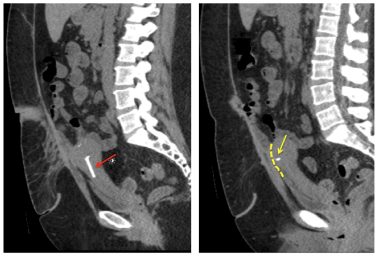
A 34-year-old gravida 4 para 4 female with obesity (body mass index 42.23 kg/m2), chronic pelvic pain, four prior cesarean sections, and Mirena intrauterine device (IUD), presented to the outpatient gynecology clinic with an acute exacerbation of pelvic pain. One month prior to presentation, she was involved in a motor vehicle accident. Immediately after, she developed new onset "poking" pain in the pelvis. Computed tomography (CT) scan of the abdomen and pelvis demonstrated the uterus positioned anteriorly in the pelvis near the anterior abdominal wall and the left arm of the IUD extending to the periphery of the anterior uterine wall (Figure 1).
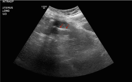
The Mirena IUD was placed nine months prior and satisfactory IUD placement was performed under anesthesia with transabdominal ultrasound guidance (Figure 2). Severe anatomic distortion of the cervix deep in the vaginal canal likely due to her habitus and adhesive disease had resulted in the inability to visualize and access the cervix on speculum examination in the office.
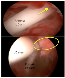
Due to ongoing pelvic pain and concern for IUD malposition, the patient elected to undergo removal of the IUD. She underwent vaginoscopic removal of the IUD under anesthesia. Despite attempts using speculums of varying sizes and lengths, the cervix could not be visualized, due to its deep and anterior position. Vaginoscopy was performed using a 5 mm rigid hysteroscope. Vaginal distension with normal saline was maintained using perineal pressure and an Allis clamp placed across the labia. The hysteroscope was advanced towards the posterior fornix to visualize the IUD strings at the cervical os. The cervical canal was easily entered without cervical dilation or use of a cervical tenaculum. The IUD stem was visualized in the lower portion of the uterine cavity and one IUD arm was noted to be pointed posteriorly, but completely visible. The other IUD arm was pointed anteriorly and penetrating the uterine wall (Figure 3). The hysteroscopic grasper was used to grasp the IUD strings. Gentle traction was used, with simultaneous removal of the hysteroscope, to remove the IUD intact.
Case 2
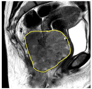
A 39-year-old gravida 2 para 2 female with a newly diagnosed rectosigmoid mass presented to the outpatient gynecology clinic desiring removal of an IUD of unknown type placed four years prior outside of the United States. The patient reported a six-month history of constipation and rectal bleeding and underwent evaluation with CT of the abdomen and pelvic demonstrating a 10 cm rectosigmoid mass (Figure 4) occupying the vaginal canal. Colonoscopy with biopsies revealed adenocarcinoma of the colon. Colorectal surgery consultation recommended further evaluation with magnetic resonance imaging (MRI) for surgical planning (Figure 5). IUD removal was recommended due to concern for susceptibility artifact and interference with image quality from the IUD during the MRI. The cervix was unable to be visualized during office speculum examination.
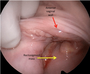
The patient was taken to the operating room and a pelvic exam under anesthesia revealed an edematous left labium majus and minus, likely due to mass effect. The cervix and IUD strings could not be palpated or visualized with a speculum. A large, firm, friable mass was appreciated along the posterior vagina, consistent with the known rectal mass (Figure 6). The rigid hysteroscope was introduced into the vagina and was able to bypass the obstruction caused by the rectal mass. Vaginoscopy allowed for visualization of the IUD strings, which were grasped allowing for intact IUD removal.
Case 3
A 59-year-old gravida 2 para 2 female presented to the outpatient gynecology clinic for evaluation of postmenopausal bleeding characterized by light spotting episodes, lasting up to two weeks in duration. Her prior gynecologic history was notable for having undergone an endometrial ablation approximately 13-years prior. A pelvic ultrasound demonstrated trace fluid in the endometrial cavity and limited visualization of the endometrium. The endometrial thickness measured approximately 0.3 to 0.7 cm. On speculum examination, the vaginal mucosa appeared atrophic. Office endometrial biopsy was attempted and the endometrial pipelle catheter was noted to encounter resistance at approximately 4-5 cm deep, suggestive of intrauterine scarring or stenosis. Pathology showed acute inflammation and rare atrophic endometrial glands.
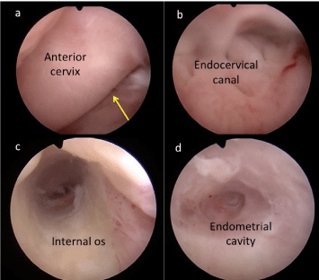
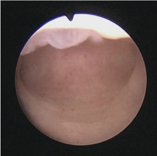
Due to the inadequate office endometrial biopsy, the patient was recommended to undergo direct cavity evaluation with hysteroscopy. A 5 mm hysteroscope was directed into the uterine cavity using a vaginoscopic approach by distending the vaginal canal with normal saline. A speculum and tenaculum were not required. The endocervical folds, or plicae palmatae, and internal cervical os served as useful anatomic landmarks during hysteroscope advancement. The cavity appeared narrow with evidence of intrauterine scarring and without focal lesions (Figure 7). Transabdominal ultrasound guidance confirmed the intrauterine placement of the hysteroscope.
Case 4
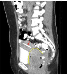
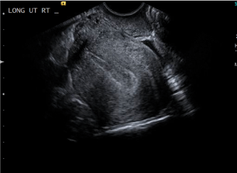
A 38-year-old gravida 2 para 2 with history of abnormal uterine bleeding was noted to have endometrial thickening of 1.2 cm and concern for possible endometrial polyp on pelvic ultrasound. The patient reported very heavy periods with passage of clots. Pelvic ultrasound was previously obtained which demonstrated thickening and vascularity of the endometrium. She continued to have abnormal uterine bleeding despite use of oral medroxyprogesterone acetate and tranexamic acid. Repeat pelvic ultrasound imaging demonstrated persistent endometrial thickening. Severe uterine retroflexion was noted on pelvic examination and confirmed by pelvic ultrasound (Figure 8). The patient elected to undergo cavity evaluation with outpatient hysteroscopy. A rigid 5 mm hysteroscope was inserted into vaginal canal. The vaginal was distended with normal saline. The hysteroscope was guided into the cervical canal. Gentle rotational maneuvers and cephalad advancement of the hysteroscope allowed for successful entry into the uterine cavity. No focal lesions or polyps were noted (Figure 9).
Discussion
Vaginoscopy is a useful, versatile, and likely underutilized surgical approach to hysteroscopy. As demonstrated in the cases explored above, vaginoscopy can also have a clear advantage over traditional hysteroscopy in patients with distorted anatomy. A vaginoscopic approach to hysteroscopy may not be used as frequently as a standard approach in many gynecological procedures due to physician unfamiliarity [14].
Within the four cases presented, we described the use of vaginoscopy to accomplish comprehensive evaluation of the genital tract and uterine cavity and other procedural goals. By performing a vaginoscopic approach in patient with class III obesity, the previously encountered difficulties were navigated with ease. Further, the difficult anatomy of a rectal mass, cervical stenosis, and a severely retroflexed uterus were all approached with vaginoscopy successfully whereas a traditional approach may have otherwise failed.
This case series and brief review of the literature aims to provide different examples in which vaginoscopy may be utilized, but this technique is by no means limited to these scenarios. In providing a step-by-step approach to vaginoscopy, as well as troubleshooting strategies, we hope that incorporating vaginoscopy in clinical practice will be less daunting for gynecologic surgeons. It is our recommendation that this technique be standard of care in outpatient hysteroscopy, as well as a reliable and reproducible technique in cases of distorted anatomy.
Data Availability
The data that support the findings of this study are available from the corresponding author upon reasonable request.
Consent
Each patient has signed an informed consent form stating that she agrees to give the authors permission to use her protected health information, with all personal identifiers removed, for scientific or educational purposes. A copy of this signed informed consent form is available upon request.
Conflict of Interest
The authors declare that they have no conflicts of interest.
Funding
No funding was received.
References
- Bettocchi S, Selvaggi L (1997) A vaginoscopic approach to reduce the pain of office hysteroscopy. The Journal of the American Association of Gynecologic Laparoscopists 4: 255-258.
- Jin L, Yi S, Wang X, et al. (2019) Application of “no-touch” hysteroscopy (vaginoscopy) for the treatment of abnormal uterine bleeding in adolescence. The Journal of Obstetrics and Gynaecology Research 45: 1913-1917.
- Ekinci S, Karnak İ, Tanyel FC, et al. (2016) Prepubertal vaginal discharge: Vaginoscopy to rule out foreign body. The Turkish Journal of Pediatrics 2016;58:168-171.
- Johary J, Xue M, Xu B, et al. (2015) Use of hysteroscope for vaginoscopy or hysteroscopy in adolescents for the diagnosis and therapeutic management of gynecologic disorders: A systematic review. Journal of Pediatric and Adolescent Gynecology 28: 29-37.
- di SpiezioSardo A, Zizolfi B, Calagna G, et al. (2016) Vaginohysteroscopy for the diagnosis and treatment of vaginal lesions. International Journal of Gynaecology and Obstetrics 133: 146-151.
- Parkin L, Sweetland S, Balkwill A, et al. (2012) Body mass index, surgery, and risk of venous thromboembolism in middle-aged women: a cohort study. Circulation 125: 1897-1904.
- Patel RS, Christie AL, Zimmern PE (2021) Role of office-based vaginoscopy in a tertiary care center. Urology Practice 9: 80-86.
- Billone V, Amorim-Costa C, Campos S, et al. (2015) Laparoscopy-like operative vaginoscopy: A new approach to manage mesh erosions. JMIG 22: 10.
- Alkhatib AA, Santoro GA, Gorgun E, et al. (2016) Tandem vaginoscopy with colonoscopy: A diagnostic technique for the assessment of colovaginal fistula. Colorectal Disease 18: 483-487.
- Smith PP, Kolhe S, O'Connor S, et al. (2019) Vaginoscopy Against Standard Treatment: A randomised controlled trial. BJOG 126: 891-899.
- de Silva PM, Carnegy A, Smith PP, et al. (2020) Vaginoscopy for office hysteroscopy: A systematic review & meta-analysis. Eur J Obstet Gynecol Reprod Biol 252: 278-285.
- (2020) The use of hysteroscopy for the diagnosis and treatment of intrauterine pathology: ACOG committee opinion, number 800. Obstetrics and Gynecology 135: e138-e148.
- Deffieux X, Gauthier T, Menager N, et al. (2014) Hysteroscopy: Guidelines for clinical practice from the French College of Gynaecologists and Obstetricians. EJOG 178: 114-122.
- Cooper NAM, Smith P, Khan KS, et al. (2010) Vaginoscopic approach to outpatient hysteroscopy: A systematic review of the effect on pain. BJOG 117: 532-239.
Corresponding Author
Sarah Swartz, MD, Department of Obstetrics and Gynecology, George Washington University Hospital, 900 23rd St. NW, Washington, DC 20037, USA
Copyright
© 2022 Swartz S. This is an open-access article distributed under the terms of the Creative Commons Attribution License, which permits unrestricted use, distribution, and reproduction in any medium, provided the original author and source are credited.