Concomitant Saline Infused Sonohysterography and Endometrial Sampling of Intrauterine Pathology Using SonoSure Device
Abstract
Background
Saline Infusion Sonohysterography (SIS) has become a routine diagnostic tool for the female reproductive tract and provides better visualization and detection of intrauterine pathology, enhancing the diagnostic accuracy of Transvaginal Ultrasound (TVS) in women with Abnormal Uterine Bleeding (AUB). While saline infusion sonohysterography and Endometrial Biopsy (EMB) are routinely used in the workup for AUB, few providers perform these procedures concurrently. We describe the feasibility and efficacy of a novel endometrial sampling device for concomitant sonohysterography and ultrasound directed sampling to evaluate abnormal uterine bleeding in thirty-three women.
Results
After SIS evaluation, the uterine cavity was adequately sampled in 91% (n = 30) of women. Pathology was consistent with traditional hysteroscopy or hysterectomy specimens, however polyps (n = 4) and leiomyomata (n = 1) were missed. It was effective and well tolerated with no post-procedural complications.
Conclusions
The SonoSure device was a practical way for a single provider to accurately assess the endometrial cavity, while maintaining patient comfort with a single cervical cannulation. Our data suggests that women with an increased risk for sampling failure - postmenopausal bleeding, history of prior failed Pipelle biopsy, and obesity, may benefit from this device. Comparison studies are underway to evaluate the diagnostic accuracy and rate of sufficient sampling for this SIS-EMB device compared to traditional sampling devices compared to hysterectomy specimens in those with AUB.
Keywords
Abnormal Uterine Bleeding, Sonohysterography, Endometrial Biopsy, SonoSure.
Background
Abnormal Uterine Bleeding (AUB), a harbinger of endometrial cancer, occurs in 20% of women between the ages of 19 to 55 [1,2]. To properly evaluate the endometrium, anatomic pathology causing AUB should be differentiated from anovulatory bleeding.
Historically, screening for endometrial cancer has been done with dilation and curettage with or without hysteroscopy. However, technological advances in instrumentation have allowed providers to sample the endometrial cavity in an outpatient setting via endometrial biopsy, suction devices, Transvaginal Ultrasound (TVS), or Saline Infusion Sonohysterography (SIS) [3]. Dilation and Curettage (D&C) and Endometrial Biopsy (EMB) curettes such as Pipelle are among the most studied techniques to evaluate the endometrium. However, the accuracy of D&C has been questioned, as 60% of these procedures sample less than half the endometrium [3]. When focal lesions are present, curettage fails to adequately sample the endometrium 38%-100% of the time [4]. On the other hand, Guido, et al. has shown that when there is global, endometrial pathology, the Pipelle device adequately sampled 97% of women with a sensitivity of 83% [5]. However, several studies have revealed that endometrial biopsy can miss a significant number of lesions [5-8]. In a meta-analysis by Dijkhuizen, et al. [9] premenopausal women were at increased risk for endometrial sampling failure compared to postmenopausal women with detection rates of 91% and 99.6% [9]. They further speculated whether the work up of perimenopausal bleeding should combine endometrial biopsy with transvaginal ultrasound to improve accuracy. Unfortunately, TVS is limited by the requirement to completely visualize the endometrial echo, which should be surrounded by an intact hypoechoic junctional zone. Often angulation of the uterus or coexisting pathology can distort the endometrium and decrease the predictive value of ultrasound imaging [10-12].
Saline infusion sonohysterography has become a routine diagnostic tool for the female reproductive tract and provides better visualization and detection of intrauterine pathology, enhancing the diagnostic accuracy of TVS in women with AUB. Moreover, it has been shown to have better predictive value in identifying endometrial pathology compared to endometrial sampling devices and to be more cost effective in comparison to dilation and curettage [13-18]. While SIS and EMB are routinely used in the workup for AUB, few providers perform these procedures concurrently - often requiring multiple office visits or multiple cervical cannulations. Here we describe the feasibility and efficacy of the SonoSure endometrial sampling device for concomitant SIS and ultrasound directed sampling of endometrial pathology for the evaluation of AUB.
Methods
From January 2014-February 2016, thirty-three pre-and postmenopausal women (30-68 years of age) undergoing evaluation for AUB were referred for SIS and directed biopsy at Arizona Gynecology Consultants, Phoenix, Arizona by KR. The indications for SIS and EMB included uterine leiomyomata and menorrhagia (n = 8), menorrhagia (n = 12), leiomyomata (n = 2), post-menopausal bleeding (n = 6), irregular menses and abnormal ultrasound findings (n = 3), and leiomyomata and infertility (n = 2). All patients provided written consent for SIS and EMB. Due to the retrospective data collection, IRB approval was not needed as per local regulations.
All patients had a documented negative urine pregnancy test and were offered to take a non-steroidal anti-inflammatory agent (Ibuprofen 400 mg to 600 mg) one hour prior to the procedure.
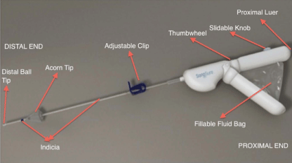
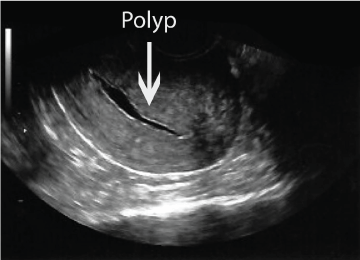
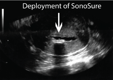
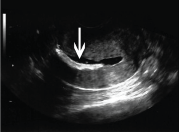
Diagnostic evaluation of the uterine cavity was performed using a disposable 3 mm malleable insertion catheter (CrossBay Medical Inc., San Diego, CA) with a built-in deployable endometrial sampling brush and a repositionable cervical sealing mechanism for uterine distension. The accommodating acorn tip fit onto the external os of the cervix and was designed not to obscure the lower uterine segment (Figure 1). Using the 50 cc re-fillable fluid injection bag on the device, 5-10 cc of sterile saline was introduced through the catheter by the operator (Figure 2a). Transvaginal sonographic images of the sagittal and transverse views of the uterus and pelvis were recorded (Voluson I, TV probe - RIC5-9w- RS - 4-9 MHz, Windows XP Embedded - 8.2.2, Zinf, Austria). When pathology was identified, and at the discretion of the physician, SIS guided extraction and/or biopsy was performed. A sliding knob on the SonoSure handle advanced the endometrial sampling brush past the distal catheter tip, into the uterine cavity at the area of interest (Figure 2b). In cases where distension media collapsed around the area of interest, the cavity was re-distended to achieve adequate visualization. After sampling was completed, the endometrial sampling brush was withdrawn from the cavity by retracting the slidable knob on the SonoSure handle in the opposite direction to withdraw the endometrial sampling brush back into the SonoSure cannula (Figure 2c). Once withdrawn, the SonoSure device was removed from the endocervix, placed within a specimen container where the endometrial sampling brush was then re-advanced to obtain cells and material for pathologic evaluation. All procedures were performed with transvaginal ultrasound guidance.
Results
The average patient age was 43.1 years of age. All 33 patients successfully underwent SIS. Twenty-nine procedures revealed submucosal filling defects, while four demonstrated a normal intrauterine cavity. In all cases, the endometrial sampling device was successfully deployed (Figure 2a, Figure 2b and Figure 2c). Three women with mild cervical stenosis required a tenaculum to allow advancement of the SonoSure device. None required cervical dilation or an assistant as a "second pair of hands". The procedure was well tolerated with the duration of 5 to 15 minutes from start to finish. There were no post-procedural complications. All patients were discharged to home within 15 minutes following the procedure.
All specimens were sent for pathologic evaluation, and thirty samples were found to be adequate. All specimens were benign. Twenty of these women underwent surgical intervention (Table 1 and Table 2). Simultaneous SIS and EMB missed four polyps and one leiomyomata. Otherwise, the surgical pathology was consistent with the samples collected with the SonoSure device. Of those with inadequate sampling (n = 3), one underwent hysteroscopy and D&C, one underwent a hysterectomy, while the other was followed clinically.
Discussion
Our case series assessed the feasibility and efficacy of concomitant SIS and ultrasound directed sampling of the endometrium. Our preliminary data suggest this SIS-EMB catheter system samples the cavity in more than 90% of cases, has a high degree of correlation with traditional hysteroscopy and hysterectomy specimens, and is well tolerated by patients. Furthermore, in post-menopausal women, all six samples revealed benign pathology consistent with hysteroscopy/hysterectomy pathology, though one missed a benign polyp, suggesting that five (82%) of the surgical procedures could have been avoided. On the other hand, we acknowledge that our results are drawn from a heterogeneous population with a small sample size (n = 33). These preliminary findings should be interpreted with caution.
The potential for directed sampling at the time of identification could reduce patient inconvenience. Endometrial sampling devices used in conjunction with SIS have been described and have been shown to be feasible, safe, and well tolerated in most patients [19]. However, perfecting ideal and inexpensive instrumentation has been a challenge. Wei, et al. previously reported the use of the Uterine Explora Curette, but 20% of cases required the need for placement of a balloon catheter caudal to Explora Curette to prevent fluid egress from the cervical canal, adding time to the procedure and making the procedure more cumbersome [20]. Authors suggested modifications to a catheter system to include an obstructive balloon system or an acorn plug to enhance the utility of this type of procedure. The SonoSure device prevents fluid egress and effectively combines SIS and EMB in the same device.
SIS has become a routine diagnostic tool for the female reproductive tract as it provides better visualization and detection of intrauterine pathology than routine TVS, has better predictive value in identifying endometrial pathology compared to endometrial sampling devices, and is more cost-effective in comparison to dilation and curettage with or without hysteroscopy [13-18]. However, even when uterine pathology is visualized with SIS, up to one third of cases obtain unsatisfactory specimens with traditional sampling devices [19,20].
Currently, the popular method to sample the endometrial cavity is with low-pressure endometrial biopsy devices such as the Pipelle. Guido, et al. has previously described the strength of this device in adequately sampling the endometrium, specifically when there is global endometrial pathology [5]. However, failure rate of Pipelle sampling procedures vary from 8%-10.4% [9,21]. Adambekov, et al. recently described the various risk factors associated with endometrial sampling failure - their findings suggest that women with postmenopausal bleeding, history of prior failed Pipelle biopsy, and obesity are at increased risk of sampling failure [22]. Perhaps these women would benefit from concomitant SIS and ultrasound directed EMB.
Conclusions
The SonoSure device was a practical way for a single provider to accurately assess the endometrial cavity, while maintaining patient comfort with a single cervical cannulation. Our data suggests that women with an increased risk for sampling failure may benefit from this device. Comparison studies are underway to evaluate the diagnostic accuracy and rate of sufficient sampling for this SIS-EMB device compared to traditional sampling devices compared to hysterectomy specimens in those with AUB.
Declaration
All of the authors had no financial support for this study. Currently, Dr. Roy serves as Chief Medical Officer and stockholder of CrossBay Medical Inc.
Authors' Contributions
KC- analyzed data, participated in writing the manuscript, KCC- planned study design, collected and analyzed data, participated in writing the manuscript, JC- planned study design, collected and analyzed data and participated in writing the manuscript, KR- planned study design, collected and analyzed data, and participated in writing the manuscript, NJ and JL- analyzed data, participated in writing the manuscript, SRL- planned study design, collected and analyzed data, participated in writing the manuscript. All authors read and approved the final manuscript.
References
- Kotdawala P, Kotdawala S, Nagar N (2013) Evaluation of endometrium in peri-menopausal abnormal uterine bleeding. J Midlife Health 4: 16-21.
- (2015) Practice Bulletin No. 149: Endometrial cancer. Obstet Gynecol 125: 1006-1026.
- Goldstein SR (2009) The role of transvaginal ultrasound or endometrial biopsy in the evaluation of the menopausal endometrium. Am J Obstet Gynecol 201: 5-11.
- Stock RJ, Kanbour A (1975) Prehysterectomy curettage. Obstet Gynecol 45: 537-541.
- Guido RS, Kanbour-Shakir A, Rulin MC, et al. (1995) Pipelle endometrial sampling. Sensitivity in the detection of endometrial cancer. J Reprod Med 40: 553-555.
- Goldschmit R, Katz Z, Blickstein I, et al. (1993) The accuracy of endometrial Pipelle sampling with and without sonographic measurement of endometrial thickness. Obstet Gynecol 82: 727-730.
- Rodriquez GC, Yaqub N, King ME (1993) A comparison of the Pipelle device and the Vabra aspirator as measured by endometrial denudation in hysterectomy specimens: the Pipelle device samples significantly less of the endometrial surface than the Vabra aspirator. Am J Obstet Gynecol 168: 55-59.
- Goldstein SR, Zeltser I, Horan CK, et al. (1997) Ultrasonography-based triage for perimenopausal patients with abnormal uterine bleeding. Am J Obstet Gynecol 177: 102-108.
- Dijkhuizen PF, Mol BW, Brolmann HA, et al. (2000) The accuracy of endometrial sampling in the diagnosis of patients with endometrial carcinoma and hyperplasia: A meta-analysis. Cancer 89: 1765-1772.
- Goldstein SR, Nachtigall M, Snyder JR, et al. (1990) Endometrial assessment by vaginal ultrasonography before endometrial sampling in patients with postmenopausal bleeding. Am J Obstet Gynecol 163: 119-123.
- Lindheim SR, Adsuar N, Kushner DM, et al. (2003) Sonohysterography: a valuable tool in evaluating the female pelvis. Obstet Gynecol Surv 58: 770-784.
- Dubinsky TJ, Parvey HR, Gormaz G, et al. (1995) Transvaginal hysterosonography: comparison with biopsy in the evaluation of postmenopausal bleeding. J Ultrasound Med 14: 887-893.
- Neele SJ, Marchien van Baal W, van der Mooren MJ, et al. (2000) Ultrasound assessment of the endometrium in healthy, asyptomatic early post-menopausal women: saline infusion sonohysterography versus transvaginal ultrasound. Ultrasound Obstet Gynecol 16: 254-259.
- Epstein E, Ramirez A, Skoog L, et al. (2001) Transvaginal sonography, saline contrast sonohysterography and hysteroscopy for the investigation of women with postmenopausal bleeding and endometrium > 5 mm. Ultrasound Obstet Gynecol 18: 157-162.
- Mathew M, Gowri V, Rizvi SG (2010) Saline infusion sonohysterography - an effective tool for evaluation of the endometrial cavity in women with abnormal uterine bleeding. Acta Obstet Gynecol Scand 89: 140-142.
- Soguktas S, Cogendez E, Kayatas SE, et al. (2012) Comparison of saline infusion sonohysterography and hysteroscopy in diagnosis of premenopausal women with abnormal uterine bleeding. Eur J Obstet Gynecol Reprod Biol 16: 66-70.
- Lindheim SR, Kavic S, Sauer MV (1999) Intraoperative Applications of Saline Infusion Ultrasonography. J Assist Reprod Genet 16: 390-394.
- Weber AM, Belinson JL, Bradley LD, et al. (1997) Vaginal ultrasonography versus endometrial biopsy in women with postmenopausal bleeding. Am J Obstet Gynecol 177: 924-929.
- Bernard JP, Metzger U, Camatte S, et al. (2002) Comparison of three catheters for endometrial sampling during sonohysterography: results of preliminary study. Journal of Obstetrics and Gynaecology 22: 84-85.
- Wei AY, Schink JC, Pritts EA, et al. (2006) Saline contrast sonohysterography and directed extraction, resection, and biopsy of intrauterine pathology using a Uterine Explora Curette. Ultrasound Obstet Gynecol 27: 202-205.
- Clark TJ, Mann CH, Shah N, et al. (2002) Accuracy of outpatient endometrial biopsy in the diagnosis of endometrial cancer: a systematic quantitative review. BJOG 109: 313-321.
- Adambekov S, Goughnour SL, Mansuria S, et al. (2017) Patient and provider factors associated with endometrial Pipelle sampling failure. Gynecol Oncol 144: 324-328.
Corresponding Author
Steven R Lindheim, MD, MMM, Department of Obstetrics and Gynecology, Boonshoft School of Medicine, Wright State University, 128 E, Apple Street, Suite 3811, Dayton, Ohio 45409, USA, Tel: 937-208-2850.
Copyright
© 2017 Cotangco K, et al. This is an open-access article distributed under the terms of the Creative Commons Attribution License, which permits unrestricted use, distribution, and reproduction in any medium, provided the original author and source are credited.

