Accessory Pulmonary Vein Posterior to the Bronchus Intermedius Encountered During Mediastinoscopy
Classifications
Mediastinoscopy, Pulmonary arteries/veins
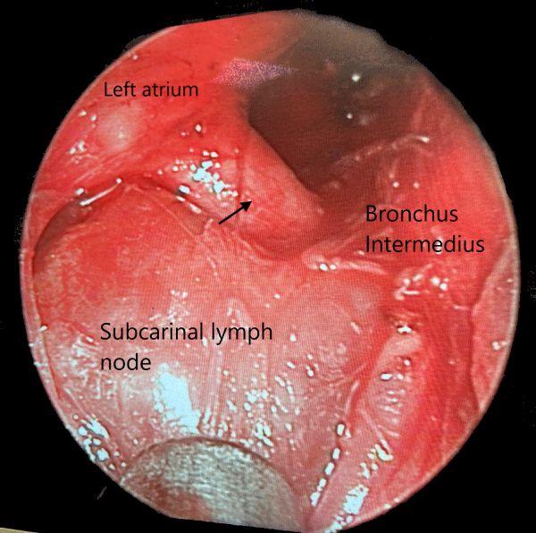
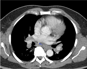
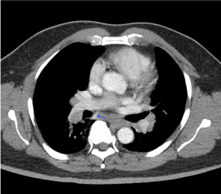
The patient was a 63-year-old, lifelong nonsmoker, Haitian man who complained for several months of early satiety and occasional epigastric pain. The patient had lost 35 pounds over the last three months. The patient2021/06/14s appetite was significantly diminished. He was referred for mediastinal and bilateral hilar adenopathy detected on CT scan. He then had a PET-CT which demonstrated numerous prominent PET-avid paratracheal, precarinal, subcarinal, aorta-pulmonary window, superior mediastinal and retroperitoneal lymph nodes. The patient then suffered an ischemic stroke. He received tPA at and recovered with a minor gait disturbance. His cardiovascular and neurological work-up did not reveal any organic disease. His initial complaints of early satiety, abdominal pain and weight loss persisted. He underwent a mediastinoscopy and lymph node biopsy. The pathology resulted as noncaseating granulomas, consistent with sarcoidosis. An accessory right pulmonary vein was observed coursing in the inferior subcarinal space and posterior to the bronchus intermedius (Figure 1, arrow). It was then retrospectively identified on the pre-operative CT scan (Figure 2 and Figure 3, arrow).
Atypical, anatomic pulmonary vein configurations are present in about a third of the population [1]. This is of significance to the thoracic surgeon, for inadvertent vascular injury can lead to life-threatening complications. Several case reports exist describing an accessory right superior pulmonary vein coursing posterior to the bronchus intermedius encountered during pulmonary lobectomy and esophagectomy [2-5]. This variation was found in 41 (5.7%) of 725 computed tomography cases, and in 9 (3.9%) of 230 right thoracotomy cases [3]. In another series, this anomaly was found in 3.9% of right thoracotomy cases [4]. This suggests that this occurrence is more prevalent than expected. Normally, no large central blood vessels pass through the subcarinal space, and the right superior pulmonary vein crosses anterior to the upper lobe bronchus. Subcarinal lymph node dissection is a common thoracic procedure and often performed during lobectomy, esophagectomy, and mediastinoscopy. Surgeons do not routinely expect there to be a large pulmonary vein in this anatomic space. Failure to recognize an anomalous vessel could result in severe bleeding and/or air embolus with suboptimal access and exposure, especially if working in the left pleural space. To the best of my knowledge this is the first reported case and in-situ photograph of an accessory right superior pulmonary vein encountered during mediastinoscopy (Figure 1). Although rare, thoracic surgeons should be aware of the possibility of anomalous pulmonary vascular anatomy and seek them out on preoperative CT scans.
References
- Porres DV, Morenza ÓP, Pallisa E, et al. (2013) Learning from the Pulmonary Veins. RadioGraphics 33: 999-1022.
- Tarazi M, Mayooran N, Philip B, et al. (2016) Anomalous right upper lobe venous drainage. J Surg Case Rep 2016.
- Asai K, Urabe N, Yajima K, et al. (2005) Right upper lobe venous drainage posterior to the bronchus intermedius: Preoperative identification by computed tomography. Ann Thorac Surg 79: 1866-1871.
- Ichiki Y, Kakizoe K, Hamatsu T, et al. (2017) A rare anomaly of the right superior pulmonary vein: Report of a case. Int J Surg Case Rep 38: 26-28.
- Matsubara T (2003) Rare but dangerous anomaly of the right pulmonary vein in subcarinal dissection. Ann Thorac Surg 75: 1026.
Corresponding Author
Erik A Sylvin, MD, JFK Medical Center, 180 JFK Drive, Suite 320, Atlantis, Florida, USA, 33462, Tel: 561-548-4900, Fax: 561-434-5165.
Copyright
© 2021 Sylvin EA. This is an open-access article distributed under the terms of the Creative Commons Attribution License, which permits unrestricted use, distribution, and reproduction in any medium, provided the original author and source are credited.