Echo-Guided Recruitment of Lung Atelectasis after Pediatric Cardiac Surgery
Abstract
Lung ultrasound is a useful tool in intensive care unit. In addition to imaging various lung and pleural diseases, LUS could guide interventional procedure. We describe a case of manual recruitment of atelectasis under lung ultrasound guidance in a child after surgical ligation of patent ductus arteriosus.
Keywords
Lung ultrasound, Congenital heart disease, Atelectasis
Introduction
Lung ultrasound (LUS) is gaining consensus in the diagnosis and management of lung disease in intensive care unit also in children [1]. Moreover, besides the imaging of various lung and pleural diseases, LUS can guide invasive or interventional procedures as thoracentesis, and needle biopsy of the pleura [2]. In this case report, we describe a manual recruitment of atelectasis under LUS in a child after surgical closure of patent ductus arteriosus.
Case Report
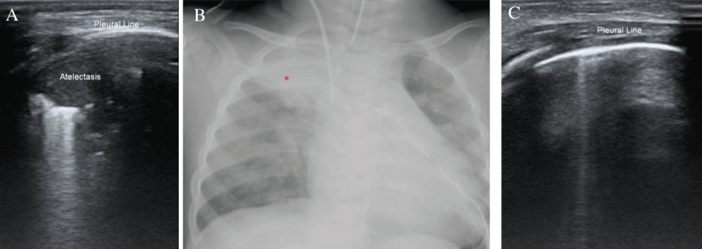
An 11-month-old female baby underwent a surgical ligation of a large patent ductus arteriosus. On first post-operative day, a right apical atelectasis was documented at both lung ultrasound (LUS) (Figure 1A) and chest X-ray (Figure 1B) while the patient was still intubated. LUS was performed with a conventional approach [1,3] using a linear probe, L12-4s, of Mindray M9 ultrasound system, Shenzhen, China. Three major scanning areas (anterior, lateral and posterior) delineated by the parasternal, anterior axillary and posterior axillary line have been identified for each hemithorax; each area has been further divided into the upper and lower half. Manual recruitment of the atelectasis under ultrasonographic guidance was attempted. After manual recruitment, LUS showed the resolution of the right apical atelectasis (Figure 1C, Video 1).
Discussion
Pulmonary complications are very common in pediatric cardiac surgery and LUS can provide findings that the clinician has to integrate with those provided by conventional radiology. In particular, LUS may allow differential diagnosis in common post-operative conditions (i.e. effusion versus atelectasis) and provide a deeper understanding in the physiology of pulmonary complications, even if focused studies are necessary. LUS also offers the advantage of being a radiation-free, easy, and cheap tool [4,5] who can be repeated whenever necessary. Thus, LUS may help to follow rapid and dynamic pulmonary changes occurring post cardiac surgery and to actively monitor invasive/non invasive manoeuvres, such as one we have described.
In conclusion, we reported an interesting application < of LUS in paediatric cardiac surgery intensive care setting.
Acknowledgments and Disclosures
The work has been supported by Italian Health Ministry Finalized Research Young Research Award 2013 Project Nr GR-2013-02358631.
References
- Daniel A Lichtenstein, Philippe Mauriat (2012) Lung ultrasound in the critically ill neonate. Curr Pediatr Rev 8: 217-223.
- Tsai TH, Yang PC (2003) Ultrasound in the diagnosis and management of pleural disease. Curr Opin Pulm Med 9: 282-290.
- Volpicelli G, Elbarbary M, Blaivas M, et al. (2012) International evidence-based recommendations for point-of-care lung ultrasound. Intensive Care Med 38: 577-591.
- Gargani L, Volpicelli G (2014) How I do it: lung ultrasound. Cardiovasc Ultrasound 12: 25.
- Cantinotti M, Giordano R, Volpicelli G, et al. (2015) Lung ultrasound in adult and paediatric cardiac surgery: is it time for routine use? Interact Cardiovasc Thorac Surg 22: 208-215.
Corresponding Author
Lamia Ait Ali, Institute of Clinical Physiology, National Research Council, Via Aurelia Sud 112, 54100 Massa, Italy, Tel: +393-40247-7742, Fax: +3905-8549-3616.
Copyright
© 2017 Cantinotti M, et al. This is an open-access article distributed under the terms of the Creative Commons Attribution License, which permits unrestricted use, distribution, and reproduction in any medium, provided the original author and source are credited.